 Movie
Movie Controller


+ Open data

- Basic information
| Entry | Database: PDB / ID: 1eiq | ||||||
|---|---|---|---|---|---|---|---|
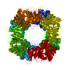
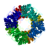
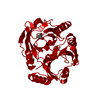
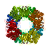
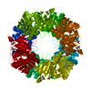
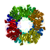
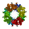
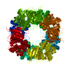
| Title | 2,3-DIHYDROXYBIPHENYL-1,2-DIOXYGENASE | ||||||
 Components Components | 2,3-DIHYDROXYBIPHENYL-1,2-DIOXYGENASE | ||||||
 Keywords Keywords | OXIDOREDUCTASE / four repetitions of beta-alpha-beta-beta-beta motifs | ||||||
| Function / homology |  Function and homology information Function and homology informationbiphenyl-2,3-diol 1,2-dioxygenase / biphenyl-2,3-diol 1,2-dioxygenase activity / xenobiotic catabolic process / ferrous iron binding Similarity search - Function | ||||||
| Biological species |  Pseudomonas sp. (bacteria) Pseudomonas sp. (bacteria) | ||||||
| Method |  X-RAY DIFFRACTION / Resolution: 2 Å X-RAY DIFFRACTION / Resolution: 2 Å | ||||||
 Authors Authors | Senda, T. | ||||||
 Citation Citation | Journal: J.Inorg.Biochem. / Year: 2001 Title: Crystal structures of substrate free and complex forms of reactivated BphC, an extradiol type ring-cleavage dioxygenase. Authors: Uragami, Y. / Senda, T. / Sugimoto, K. / Sato, N. / Nagarajan, V. / Masai, E. / Fukuda, M. / Mitsu, Y. #1: Journal: PROC.JPN.ACAD.,SER.B / Year: 1995Title: Three-dimensional Structure of 2,3-dihydroxybiphenyl dioxygenase (BphC enzyme) from Pseudomonas sp. strain KKS102 having Polychlorinated Biphenyl (PCB) Degrading Activity. Authors: Sugiyama, K. / Senda, T. / Kimbara, K. / Fukuda, M. / Mitsui, Y. | ||||||
| History |
|
- Structure visualization
| Structure viewer | Molecule: MolmilJmol/JSmol |
|---|
- Downloads & links
-Download
| PDBx/mmCIF format | 1eiq.cif.gz | 71.7 KB | Display | PDBx/mmCIF format |
|---|---|---|---|---|
| PDB format | pdb1eiq.ent.gz | 53.5 KB | Display | PDB format |
| PDBx/mmJSON format | 1eiq.json.gz | Tree view | PDBx/mmJSON format | |
| Others |  Other downloads Other downloads |
-Validation report
| Arichive directory | https://data.pdbj.org/pub/pdb/validation_reports/ei/1eiqftp://data.pdbj.org/pub/pdb/validation_reports/ei/1eiq | HTTPS FTP |
|---|
-Related structure data


-Links
 PDBj
PDBj- Assembly
| Deposited unit | 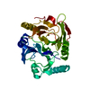
| ||||||||||||
|---|---|---|---|---|---|---|---|---|---|---|---|---|---|
| 1 | x 8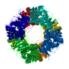
| ||||||||||||
| Unit cell |
| ||||||||||||
| Components on special symmetry positions |
|
-Components
| #1: Protein | Mass: 32151.545 Da / Num. of mol.: 1 Source method: isolated from a genetically manipulated source Source: (gene. exp.) Pseudomonas sp. (bacteria) / Strain: KKS102 / Plasmid: PHNA2 / Production host: References: UniProt: P17297, biphenyl-2,3-diol 1,2-dioxygenase |
|---|---|
| #2: Chemical | ChemComp-FE /   Mass: 55.845 Da / Num. of mol.: 1 / Source method: obtained synthetically / Formula: Fe Mass: 55.845 Da / Num. of mol.: 1 / Source method: obtained synthetically / Formula: Fe |
| #3: Water | ChemComp-HOH /  Mass: 18.015 Da / Num. of mol.: 174 / Source method: isolated from a natural source / Formula: H2O Mass: 18.015 Da / Num. of mol.: 174 / Source method: isolated from a natural source / Formula: H2O |
| Compound details | Substrate free form. Structure determined under aerobic condition (inactive form structure) |
-Experimental details
-Experiment
| Experiment | Method: X-RAY DIFFRACTION / Number of used crystals: 1 |
|---|
- Sample preparation
| Crystal | Density Matthews: 3.14 Å3/Da / Density % sol: 60.84 % | |||||||||||||||||||||||||
|---|---|---|---|---|---|---|---|---|---|---|---|---|---|---|---|---|---|---|---|---|---|---|---|---|---|---|
| Crystal grow | Temperature: 285 K / pH: 7.5 Details: 285K, Tris/HCl, Ammonium sulfate, hexylene glycol, pH 7.5 | |||||||||||||||||||||||||
| Crystal grow | *PLUS Temperature: 12 ℃ / Method: batch method | |||||||||||||||||||||||||
| Components of the solutions | *PLUS
|
-Data collection
| Diffraction | Mean temperature: 100 K |
|---|---|
| Diffraction source | Source: ROTATING ANODE / Type: RIGAKU RU200 / Wavelength: 1.542 |
| Detector | Type: RIGAKU RAXIS IIC / Detector: IMAGE PLATE / Date: Oct 28, 1998 |
| Radiation | Protocol: SINGLE WAVELENGTH / Monochromatic (M) / Laue (L): M / Scattering type: x-ray |
| Radiation wavelength | Wavelength: 1.542 Å / Relative weight: 1 |
| Reflection | Resolution: 1.98→60 Å / Num. all: 27236 / Num. obs: 25056 / % possible obs: 92 % / Observed criterion σ(F): 0 / Observed criterion σ(I): 1 / Redundancy: 2.6 % / Biso Wilson estimate: 34.9 Å2 / Rmerge(I) obs: 0.082 / Net I/σ(I): 11.5 |
| Reflection shell | Resolution: 1.98→2.25 Å / Redundancy: 1.6 % / Rmerge(I) obs: 0.201 / % possible all: 83.5 |
| Reflection | *PLUS Highest resolution: 2 Å / Num. obs: 24620 / % possible obs: 88.1 % |
| Reflection shell | *PLUS % possible obs: 83.5 % |
- Processing
| Software |
| ||||||||||||||||||||
|---|---|---|---|---|---|---|---|---|---|---|---|---|---|---|---|---|---|---|---|---|---|
| Refinement | Resolution: 2→60 Å / σ(F): 1 / σ(I): 0 / Stereochemistry target values: Engh & Huber
| ||||||||||||||||||||
| Refinement step | Cycle: LAST / Resolution: 2→60 Å
| ||||||||||||||||||||
| Refine LS restraints |
| ||||||||||||||||||||
| Software | *PLUS Name: X-PLOR / Version: 3.851 / Classification: refinement | ||||||||||||||||||||
| Refinement | *PLUS Num. reflection all: 24620 / Rfactor Rfree: 0.234 / Rfactor Rwork: 0.194 | ||||||||||||||||||||
| Solvent computation | *PLUS | ||||||||||||||||||||
| Displacement parameters | *PLUS | ||||||||||||||||||||
| Refine LS restraints | *PLUS
|
