 Movie
Movie Controller

[English] 日本語
 Yorodumi
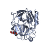
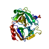
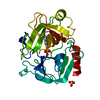
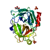
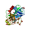
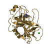
- PDB-3sbk: Russell's viper venom serine proteinase, RVV-V (PPACK-bound form) -
+ Open data

- Basic information
| Entry | Database: PDB / ID: 3sbk | ||||||
|---|---|---|---|---|---|---|---|
| Title | Russell's viper venom serine proteinase, RVV-V (PPACK-bound form) | ||||||
 Components Components | Vipera russelli proteinase RVV-V gamma | ||||||
 Keywords Keywords | HYDROLASE/HYDROLASE INHIBITOR / serine proteinase / glycosylation / double six-stranded beta-barrels / Hydrolase / HYDROLASE-HYDROLASE INHIBITOR complex | ||||||
| Function / homology |  Function and homology information Function and homology informationsnake venom factor V activator / secretory granule / toxin activity / serine-type endopeptidase activity / proteolysis / extracellular region Similarity search - Function | ||||||
| Biological species |  Daboia russellii siamensis (Siamese Russell's viper) Daboia russellii siamensis (Siamese Russell's viper) | ||||||
| Method |  X-RAY DIFFRACTION / SYNCHROTRON / MOLECULAR REPLACEMENT / Resolution: 2.55 Å X-RAY DIFFRACTION / SYNCHROTRON / MOLECULAR REPLACEMENT / Resolution: 2.55 Å | ||||||
 Authors Authors | Nakayama, D. / Ben Ammar, Y. / Takeda, S. | ||||||
 Citation Citation | Journal: Febs Lett. / Year: 2011 Title: Structural basis of coagulation factor V recognition for cleavage by RVV-V Authors: Nakayama, D. / Ben Ammar, Y. / Miyata, T. / Takeda, S. #1: Journal: Acta Crystallogr.,Sect.F / Year: 2009 Title: Crystallization and preliminary X-ray crystallographic analysis of blood coagulation factor V-activating proteinase (RVV-V) from Russell's viper venom Authors: Nakayama, D. / Ben Ammar, Y. / Takeda, S. | ||||||
| History |
|
- Structure visualization
| Structure viewer | Molecule: MolmilJmol/JSmol |
|---|
- Downloads & links
-Download
| PDBx/mmCIF format | 3sbk.cif.gz | 61.7 KB | Display | PDBx/mmCIF format |
|---|---|---|---|---|
| PDB format | pdb3sbk.ent.gz | 44.1 KB | Display | PDB format |
| PDBx/mmJSON format | 3sbk.json.gz | Tree view | PDBx/mmJSON format | |
| Others |  Other downloads Other downloads |
-Validation report
| Arichive directory | https://data.pdbj.org/pub/pdb/validation_reports/sb/3sbkftp://data.pdbj.org/pub/pdb/validation_reports/sb/3sbk | HTTPS FTP |
|---|
-Related structure data
| Related structure data |  3s9aSC  3s9bC  3s9cC S: Starting model for refinement C: citing same article ( |
|---|---|
| Similar structure data |


-Links
 PDBj
PDBj

- Assembly
| Deposited unit | 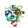
| ||||||||
|---|---|---|---|---|---|---|---|---|---|
| 1 |
| ||||||||
| Unit cell |
|
-Components
| #1: Protein | Mass: 25960.971 Da / Num. of mol.: 1 / Source method: isolated from a natural source Source: (natural) Daboia russellii siamensis (Siamese Russell's viper)Tissue: venom / References: UniProt: P18965, snake venom factor V activator |
|---|---|
| #2: Sugar | ChemComp-NAG /   Type: D-saccharide, beta linking / Mass: 221.208 Da / Num. of mol.: 1 Type: D-saccharide, beta linking / Mass: 221.208 Da / Num. of mol.: 1Source method: isolated from a genetically manipulated source Formula: C8H15NO6 |
| #3: Chemical | ChemComp-0G6 /   Type: peptide-like, Peptide-like / Class: Inhibitor / Mass: 453.986 Da / Num. of mol.: 1 / Source method: obtained synthetically / Formula: C21H34ClN6O3 / References: D-Phe-Pro-Arg-CH2Cl Type: peptide-like, Peptide-like / Class: Inhibitor / Mass: 453.986 Da / Num. of mol.: 1 / Source method: obtained synthetically / Formula: C21H34ClN6O3 / References: D-Phe-Pro-Arg-CH2Cl |
| #4: Water | ChemComp-HOH /  Mass: 18.015 Da / Num. of mol.: 17 / Source method: isolated from a natural source / Formula: H2O Mass: 18.015 Da / Num. of mol.: 17 / Source method: isolated from a natural source / Formula: H2O |
| Has protein modification | Y |
| Sequence details | THE RESIDUE NUMBERING IS NOT SEQUENTIAL. THE RESIDUE NUMBERING IS BASED ON THE TOPOLOGICAL ...THE RESIDUE NUMBERING IS NOT SEQUENTIAL |
-Experimental details
-Experiment
| Experiment | Method: X-RAY DIFFRACTION / Number of used crystals: 1 |
|---|
- Sample preparation
| Crystal | Density Matthews: 2.79 Å3/Da / Density % sol: 55.96 % |
|---|---|
| Crystal grow | Temperature: 293 K / Method: vapor diffusion, sitting drop / pH: 7 Details: 9.6% PEG 3350, 0.8% tryptone, 40mM Na/HEPES , pH 7.0, VAPOR DIFFUSION, SITTING DROP, temperature 293K |
-Data collection
| Diffraction | Mean temperature: 100 K |
|---|---|
| Diffraction source | Source: SYNCHROTRON / Site: SPring-8  / Beamline: BL41XU / Wavelength: 1 Å / Beamline: BL41XU / Wavelength: 1 Å |
| Detector | Type: RAYONIX MX-225 / Detector: CCD / Date: Jul 14, 2009 / Details: mirrors |
| Radiation | Monochromator: Rotated-inclined double-crystal / Protocol: SINGLE WAVELENGTH / Monochromatic (M) / Laue (L): M / Scattering type: x-ray |
| Radiation wavelength | Wavelength: 1 Å / Relative weight: 1 |
| Reflection | Resolution: 2.55→50 Å / Num. all: 10342 / Num. obs: 10311 / % possible obs: 99.7 % / Observed criterion σ(F): 0 / Observed criterion σ(I): 0 / Redundancy: 20.4 % / Rmerge(I) obs: 0.065 / Net I/σ(I): 33.8 |
| Reflection shell | Resolution: 2.55→2.64 Å / Redundancy: 21.4 % / Rmerge(I) obs: 0.267 / Mean I/σ(I) obs: 15.8 / Num. unique all: 993 / % possible all: 100 |
- Processing
| Software |
| |||||||||||||||||||||||||||||||||||||||||||||||||||||||||||||||||
|---|---|---|---|---|---|---|---|---|---|---|---|---|---|---|---|---|---|---|---|---|---|---|---|---|---|---|---|---|---|---|---|---|---|---|---|---|---|---|---|---|---|---|---|---|---|---|---|---|---|---|---|---|---|---|---|---|---|---|---|---|---|---|---|---|---|---|
| Refinement | Method to determine structure: MOLECULAR REPLACEMENT Starting model: PDB ENTRY 3S9A Resolution: 2.55→28.74 Å / Cor.coef. Fo:Fc: 0.924 / Cor.coef. Fo:Fc free: 0.852 / SU B: 14.957 / SU ML: 0.318 / Cross valid method: THROUGHOUT / ESU R: 0.584 / ESU R Free: 0.373 / Stereochemistry target values: MAXIMUM LIKELIHOOD
| |||||||||||||||||||||||||||||||||||||||||||||||||||||||||||||||||
| Solvent computation | Ion probe radii: 0.8 Å / Shrinkage radii: 0.8 Å / VDW probe radii: 1.4 Å / Solvent model: MASK | |||||||||||||||||||||||||||||||||||||||||||||||||||||||||||||||||
| Displacement parameters | Biso mean: 52.2571 Å2
| |||||||||||||||||||||||||||||||||||||||||||||||||||||||||||||||||
| Refinement step | Cycle: LAST / Resolution: 2.55→28.74 Å
| |||||||||||||||||||||||||||||||||||||||||||||||||||||||||||||||||
| Refine LS restraints |
| |||||||||||||||||||||||||||||||||||||||||||||||||||||||||||||||||
| LS refinement shell | Resolution: 2.551→2.617 Å / Total num. of bins used: 20
|
