 Movie
Movie Controller

[English] 日本語
 Yorodumi
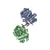
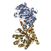
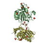
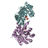
- PDB-3rp2: THE STRUCTURE OF RAT MAST CELL PROTEASE II AT 1.9-ANGSTROMS RESOLUTION -
+ Open data

- Basic information
| Entry | Database: PDB / ID: 3rp2 | ||||||
|---|---|---|---|---|---|---|---|
| Title | THE STRUCTURE OF RAT MAST CELL PROTEASE II AT 1.9-ANGSTROMS RESOLUTION | ||||||
 Components Components | RAT MAST CELL PROTEASE II | ||||||
 Keywords Keywords | SERINE PROTEINASE | ||||||
| Function / homology |  Function and homology information Function and homology informationHydrolases; Acting on peptide bonds (peptidases); Serine endopeptidases / protein maturation / serine-type endopeptidase activity / proteolysis / : Similarity search - Function | ||||||
| Biological species |  | ||||||
| Method |  X-RAY DIFFRACTION / Resolution: 1.9 Å X-RAY DIFFRACTION / Resolution: 1.9 Å | ||||||
 Authors Authors | Reynolds, R. / Remington, S. / Weaver, L. / Fischer, R. / Anderson, W. / Ammon, H. / Matthews, B. | ||||||
 Citation Citation | Journal: Biochemistry / Year: 1988 Title: The structure of rat mast cell protease II at 1.9-A resolution. Authors: Remington, S.J. / Woodbury, R.G. / Reynolds, R.A. / Matthews, B.W. / Neurath, H. #1: Journal: Acta Crystallogr.,Sect.B / Year: 1985Title: Structure of a Serine Protease from Rat Mast Cells Determined from Twinned Crystals by Isomorphous and Molecular Replacement Authors: Reynolds, R.A. / Remington, S.J. / Weaver, L.H. / Fisher, R.G. / Anderson, W.F. / Ammon, H.L. / Matthews, B.W. #2: Journal: Biochemistry / Year: 1978Title: Covalent Structure of a Group-Specific Protease from Rat Small Intestine Authors: Woodbury, R.G. / Katunuma, N. / Kobayashi, K. / Titani, K. / Neurath, H. #3: Journal: Biochemistry / Year: 1978Title: Covalent Structure of a Group-Specific Protease from Rat Small Intestine. Appendix. Crystallographic Data for a Group Specific Protease from Rat Intestine Authors: Anderson, W.F. / Matthews, B.W. / Woodbury, R.G. #4: Journal: Proc.Natl.Acad.Sci.USA / Year: 1978Title: Immunofluorescent Localization of a Serine Protease in Rat Small Intestine Authors: Woodbury, R.G. / Gruzenski, G.M. / Lagunoff, D. | ||||||
| History |
|
- Structure visualization
| Structure viewer | Molecule: MolmilJmol/JSmol |
|---|
- Downloads & links
-Download
| PDBx/mmCIF format | 3rp2.cif.gz | 101.6 KB | Display | PDBx/mmCIF format |
|---|---|---|---|---|
| PDB format | pdb3rp2.ent.gz | 75.4 KB | Display | PDB format |
| PDBx/mmJSON format | 3rp2.json.gz | Tree view | PDBx/mmJSON format | |
| Others |  Other downloads Other downloads |
-Validation report
| Arichive directory | https://data.pdbj.org/pub/pdb/validation_reports/rp/3rp2ftp://data.pdbj.org/pub/pdb/validation_reports/rp/3rp2 | HTTPS FTP |
|---|
-Related structure data
| Similar structure data |
|---|


-Links
 PDBj
PDBj- Assembly
| Deposited unit | 
| ||||||||
|---|---|---|---|---|---|---|---|---|---|
| 1 |
| ||||||||
| Unit cell |
| ||||||||
| Atom site foot note | 1: RESIDUES PRO A 225 AND PRO B 225 ARE CIS-PROLINES. 2: AN OCCUPANCY OF 0.0 INDICATES THAT THE COORDINATES WERE GENERATED USING STEREOCHEMICAL CRITERIA. | ||||||||
| Noncrystallographic symmetry (NCS) | NCS oper: (Code: given Matrix: (0.9955, 0.0465, -0.08263), Vector: Details | THE TRANSFORMATION GIVEN ON THE *MTRIX* RECORDS BELOW WILL YIELD APPROXIMATE COORDINATES FOR THE B CHAIN WHEN APPLIED TO THE A CHAIN. | |
-Components
| #1: Protein | Mass: 24802.816 Da / Num. of mol.: 2 Source method: isolated from a genetically manipulated source Source: (gene. exp.) #2: Water | ChemComp-HOH / |  Mass: 18.015 Da / Num. of mol.: 123 / Source method: isolated from a natural source / Formula: H2O Mass: 18.015 Da / Num. of mol.: 123 / Source method: isolated from a natural source / Formula: H2OCompound details | RMCPII HAS ALSO BEEN REFERRED TO AS GSP (GROUP SPECIFIC PROTEASE) IN OLDER LITERATURE | Has protein modification | Y | |
|---|
-Experimental details
-Experiment
| Experiment | Method: X-RAY DIFFRACTION |
|---|
- Sample preparation
| Crystal | Density Matthews: 3.44 Å3/Da / Density % sol: 64.27 % |
|---|---|
| Crystal grow | *PLUS Method: unknown |
| Components of the solutions | *PLUS Conc.: 23 %sat / Common name: ammonium sulfate |
-Data collection
| Reflection | *PLUS |
|---|
- Processing
| Software | Name: EREF / Classification: refinement | ||||||||||||||||||||||||||||||||||||||||||||||||||||||||||||
|---|---|---|---|---|---|---|---|---|---|---|---|---|---|---|---|---|---|---|---|---|---|---|---|---|---|---|---|---|---|---|---|---|---|---|---|---|---|---|---|---|---|---|---|---|---|---|---|---|---|---|---|---|---|---|---|---|---|---|---|---|---|
| Refinement | Resolution: 1.9→5 Å Details: RESIDUE 237 OF EACH CHAIN IS TRP. IN THE PAPER CITED AS REFERENCE 2 ABOVE RESIDUE 237 WAS IDENTIFIED AS THR BUT THIS WAS A TYPOGRAPHICAL ERROR IN THE PAPER.
| ||||||||||||||||||||||||||||||||||||||||||||||||||||||||||||
| Refinement step | Cycle: LAST / Resolution: 1.9→5 Å
| ||||||||||||||||||||||||||||||||||||||||||||||||||||||||||||
| Refine LS restraints |
| ||||||||||||||||||||||||||||||||||||||||||||||||||||||||||||
| Refinement | *PLUS Highest resolution: 1.9 Å / Lowest resolution: 5 Å / Num. reflection obs: 28782 / Rfactor obs: 0.191 | ||||||||||||||||||||||||||||||||||||||||||||||||||||||||||||
| Solvent computation | *PLUS | ||||||||||||||||||||||||||||||||||||||||||||||||||||||||||||
| Displacement parameters | *PLUS | ||||||||||||||||||||||||||||||||||||||||||||||||||||||||||||
| Refine LS restraints | *PLUS Type: o_angle_d |
