 Movie
Movie Controller

[English] 日本語
 Yorodumi
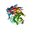
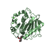
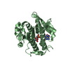
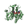
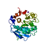
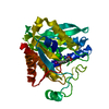
- PDB-3vis: Crystal structure of cutinase Est119 from Thermobifida alba AHK119 -
+ Open data

- Basic information
| Entry | Database: PDB / ID: 3vis | ||||||
|---|---|---|---|---|---|---|---|
| Title | Crystal structure of cutinase Est119 from Thermobifida alba AHK119 | ||||||
 Components Components | Esterase | ||||||
 Keywords Keywords | HYDROLASE / alpha/beta-hydrolase fold / esterase / polyethylene terephthalate | ||||||
| Function / homology |  Function and homology information Function and homology informationpoly(ethylene terephthalate) hydrolase / cutinase / cutinase activity / periplasmic space / extracellular region / metal ion binding Similarity search - Function | ||||||
| Biological species |   Thermobifida alba (bacteria) Thermobifida alba (bacteria) | ||||||
| Method |  X-RAY DIFFRACTION / SYNCHROTRON / SAD, molecular replacement / Resolution: 1.76 Å X-RAY DIFFRACTION / SYNCHROTRON / SAD, molecular replacement / Resolution: 1.76 Å | ||||||
 Authors Authors | Kitadokoro, K. / Thumarat, U. / Nakamura, R. / Nishimura, K. / Karatani, H. / Suzuki, H. / Kawai, F. | ||||||
 Citation Citation | Journal: POLYM.DEGRAD.STAB. / Year: 2012 Title: Crystal structure of cutinase Est119 from Thermobida alba AHK119 that can degrade modpolyethylene terephthalate at 1.76 A resolution. Authors: Kitadokoro, K. / Thumarat, U. / Nakamura, R. / Nishimura, K. / Karatani, H. / Suzuki, H. / Kawai, F. | ||||||
| History |
|
- Structure visualization
| Structure viewer | Molecule: MolmilJmol/JSmol |
|---|
- Downloads & links
-Download
| PDBx/mmCIF format | 3vis.cif.gz | 216.2 KB | Display | PDBx/mmCIF format |
|---|---|---|---|---|
| PDB format | pdb3vis.ent.gz | 173.1 KB | Display | PDB format |
| PDBx/mmJSON format | 3vis.json.gz | Tree view | PDBx/mmJSON format | |
| Others |  Other downloads Other downloads |
-Validation report
| Arichive directory | https://data.pdbj.org/pub/pdb/validation_reports/vi/3visftp://data.pdbj.org/pub/pdb/validation_reports/vi/3vis | HTTPS FTP |
|---|
-Related structure data
| Similar structure data |
|---|


-Links
 PDBj
PDBj
- Assembly
| Deposited unit | 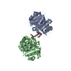
| ||||||||
|---|---|---|---|---|---|---|---|---|---|
| 1 | 
| ||||||||
| 2 | 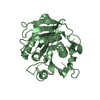
| ||||||||
| Unit cell |
|
-Components
| #1: Protein | Mass: 33215.188 Da / Num. of mol.: 2 Source method: isolated from a genetically manipulated source Source: (gene. exp.) Thermobifida alba (bacteria) / Strain: AHK119 / Gene: est2 / Plasmid: pQE80L-est119 / Production host: #2: Chemical | ChemComp-PE4 / |   Mass: 354.436 Da / Num. of mol.: 1 / Source method: obtained synthetically / Formula: C16H34O8 / Comment: precipitant*YM Mass: 354.436 Da / Num. of mol.: 1 / Source method: obtained synthetically / Formula: C16H34O8 / Comment: precipitant*YM#3: Water | ChemComp-HOH / |  Mass: 18.015 Da / Num. of mol.: 454 / Source method: isolated from a natural source / Formula: H2O Mass: 18.015 Da / Num. of mol.: 454 / Source method: isolated from a natural source / Formula: H2OHas protein modification | Y | |
|---|
-Experimental details
-Experiment
| Experiment | Method: X-RAY DIFFRACTION / Number of used crystals: 1 |
|---|
- Sample preparation
| Crystal grow | Temperature: 293 K / Method: vapor diffusion, sitting drop / pH: 6.2 Details: 25% (w/v) PEG 1000, 0.2M sodium chloride, 0.1M sodium potassium phosphate, pH 6.2, VAPOR DIFFUSION, SITTING DROP, temperature 293K |
|---|
-Data collection
| Diffraction | Mean temperature: 100 K |
|---|---|
| Diffraction source | Source: SYNCHROTRON / Site: SPring-8  / Beamline: BL44XU / Wavelength: 0.9 Å / Beamline: BL44XU / Wavelength: 0.9 Å |
| Detector | Type: RAYONIX MX225HE / Detector: CCD / Date: Jun 24, 2011 / Details: mirrors |
| Radiation | Monochromator: GRAPHITE / Protocol: SINGLE WAVELENGTH / Monochromatic (M) / Laue (L): M / Scattering type: x-ray |
| Radiation wavelength | Wavelength: 0.9 Å / Relative weight: 1 |
| Reflection | Resolution: 1.76→56.8 Å / Num. all: 44072 / Num. obs: 41770 / % possible obs: 96.9 % / Observed criterion σ(F): 1 / Observed criterion σ(I): 1 / Redundancy: 3.3 % / Biso Wilson estimate: 17 Å2 / Rmerge(I) obs: 0.099 |
| Reflection shell | Resolution: 1.76→1.82 Å / % possible all: 98 |
- Processing
| Software |
| |||||||||||||||||||||||||||||||||||||||||||||||||||||||||||||||||||||||||||
|---|---|---|---|---|---|---|---|---|---|---|---|---|---|---|---|---|---|---|---|---|---|---|---|---|---|---|---|---|---|---|---|---|---|---|---|---|---|---|---|---|---|---|---|---|---|---|---|---|---|---|---|---|---|---|---|---|---|---|---|---|---|---|---|---|---|---|---|---|---|---|---|---|---|---|---|---|
| Refinement | Method to determine structure: SAD, molecular replacement / Resolution: 1.76→56.75 Å / Cor.coef. Fo:Fc: 0.968 / Cor.coef. Fo:Fc free: 0.947 / SU B: 4.559 / SU ML: 0.073 / Cross valid method: THROUGHOUT / σ(F): 1 / σ(I): 1 / ESU R Free: 0.117 / Stereochemistry target values: MAXIMUM LIKELIHOOD
| |||||||||||||||||||||||||||||||||||||||||||||||||||||||||||||||||||||||||||
| Solvent computation | Ion probe radii: 0.8 Å / Shrinkage radii: 0.8 Å / VDW probe radii: 1.2 Å / Solvent model: MASK | |||||||||||||||||||||||||||||||||||||||||||||||||||||||||||||||||||||||||||
| Displacement parameters | Biso mean: 21.825 Å2
| |||||||||||||||||||||||||||||||||||||||||||||||||||||||||||||||||||||||||||
| Refinement step | Cycle: LAST / Resolution: 1.76→56.75 Å
| |||||||||||||||||||||||||||||||||||||||||||||||||||||||||||||||||||||||||||
| Refine LS restraints |
| |||||||||||||||||||||||||||||||||||||||||||||||||||||||||||||||||||||||||||
| LS refinement shell | Resolution: 1.762→1.808 Å / Total num. of bins used: 20
| |||||||||||||||||||||||||||||||||||||||||||||||||||||||||||||||||||||||||||
| Refinement TLS params. | Method: refined / Refine-ID: X-RAY DIFFRACTION
| |||||||||||||||||||||||||||||||||||||||||||||||||||||||||||||||||||||||||||
| Refinement TLS group |
|
