 Movie
Movie Controller

[English] 日本語
 Yorodumi
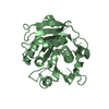
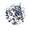
- PDB-5lul: Structure of a triple variant of cutinase 2 from Thermobifida cel... -
+ Open data

- Basic information
| Entry | Database: PDB / ID: 5lul | |||||||||||||||||||||
|---|---|---|---|---|---|---|---|---|---|---|---|---|---|---|---|---|---|---|---|---|---|---|
| Title | Structure of a triple variant of cutinase 2 from Thermobifida cellulosilytica | |||||||||||||||||||||
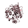
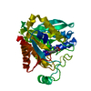
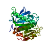
 Components Components | Cutinase 2 | |||||||||||||||||||||
 Keywords Keywords | HYDROLASE / cutinase / alpha/beta hydrolase / poly(ethyleneterephthlate) (PET) | |||||||||||||||||||||
| Function / homology |  Function and homology information Function and homology informationpoly(ethylene terephthalate) hydrolase / cutinase / cutinase activity / periplasmic space / extracellular region Similarity search - Function | |||||||||||||||||||||
| Biological species |   Thermobifida cellulosilytica (bacteria) Thermobifida cellulosilytica (bacteria) | |||||||||||||||||||||
| Method |  X-RAY DIFFRACTION / SYNCHROTRON / MOLECULAR REPLACEMENT / Resolution: 1.9 Å X-RAY DIFFRACTION / SYNCHROTRON / MOLECULAR REPLACEMENT / Resolution: 1.9 Å | |||||||||||||||||||||
 Authors Authors | Hromic, A. / Lyskowski, A. / Gruber, K. | |||||||||||||||||||||
| Funding support |  Austria, 6items Austria, 6items
| |||||||||||||||||||||
 Citation Citation | Journal: Biotechnol. Bioeng. / Year: 2017 Title: Small cause, large effect: Structural characterization of cutinases from Thermobifida cellulosilytica. Authors: Ribitsch, D. / Hromic, A. / Zitzenbacher, S. / Zartl, B. / Gamerith, C. / Pellis, A. / Jungbauer, A. / yskowski, A. / Steinkellner, G. / Gruber, K. / Tscheliessnig, R. / Herrero Acero, E. / Guebitz, G.M. | |||||||||||||||||||||
| History |
|
- Structure visualization
| Structure viewer | Molecule: MolmilJmol/JSmol |
|---|
- Downloads & links
-Download
| PDBx/mmCIF format | 5lul.cif.gz | 120.8 KB | Display | PDBx/mmCIF format |
|---|---|---|---|---|
| PDB format | pdb5lul.ent.gz | 93 KB | Display | PDB format |
| PDBx/mmJSON format | 5lul.json.gz | Tree view | PDBx/mmJSON format | |
| Others |  Other downloads Other downloads |
-Validation report
| Arichive directory | https://data.pdbj.org/pub/pdb/validation_reports/lu/5lulftp://data.pdbj.org/pub/pdb/validation_reports/lu/5lul | HTTPS FTP |
|---|
-Related structure data
| Related structure data |  5luiC 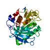 5lujC 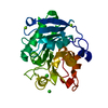 5lukC  1jfrS S: Starting model for refinement C: citing same article ( |
|---|---|
| Similar structure data |


-Links
 PDBj
PDBj
- Assembly
| Deposited unit | 
| ||||||||||||
|---|---|---|---|---|---|---|---|---|---|---|---|---|---|
| 1 | 
| ||||||||||||
| 2 | 
| ||||||||||||
| Unit cell |
| ||||||||||||
| Components on special symmetry positions |
|
-Components
| #1: Protein | Mass: 28787.189 Da / Num. of mol.: 2 / Mutation: R19S, R29N, A30V Source method: isolated from a genetically manipulated source Source: (gene. exp.) Thermobifida cellulosilytica (bacteria)Gene: cut2 / Production host: #2: Chemical | ChemComp-CA / |   Mass: 40.078 Da / Num. of mol.: 1 / Source method: obtained synthetically / Formula: Ca Mass: 40.078 Da / Num. of mol.: 1 / Source method: obtained synthetically / Formula: Ca#3: Chemical | ChemComp-CL / |   Mass: 35.453 Da / Num. of mol.: 1 / Source method: obtained synthetically / Formula: Cl Mass: 35.453 Da / Num. of mol.: 1 / Source method: obtained synthetically / Formula: Cl#4: Water | ChemComp-HOH / |  Mass: 18.015 Da / Num. of mol.: 379 / Source method: isolated from a natural source / Formula: H2O Mass: 18.015 Da / Num. of mol.: 379 / Source method: isolated from a natural source / Formula: H2OHas protein modification | Y | |
|---|
-Experimental details
-Experiment
| Experiment | Method: X-RAY DIFFRACTION / Number of used crystals: 1 |
|---|
- Sample preparation
| Crystal | Density Matthews: 2.3 Å3/Da / Density % sol: 46.58 % |
|---|---|
| Crystal grow | Temperature: 293.15 K / Method: vapor diffusion Details: 12.5% w/v PEG 1000, 12.5% w/v PEG 3350, 12.5% v/v MPD, 0.02 M of each carboxylic acid (0.2 M sodium formate, 0.2 M ammonium acetate, 0.2 M trisodium citrate, 0.2 M sodium potassium L- ...Details: 12.5% w/v PEG 1000, 12.5% w/v PEG 3350, 12.5% v/v MPD, 0.02 M of each carboxylic acid (0.2 M sodium formate, 0.2 M ammonium acetate, 0.2 M trisodium citrate, 0.2 M sodium potassium L-tartrate, 0.2 M sodium oxamate) and 0.1 M MOPS/HEPES-Na pH 7.5. |
-Data collection
| Diffraction | Mean temperature: 100 K |
|---|---|
| Diffraction source | Source: SYNCHROTRON / Site: ESRF  / Beamline: BM30A / Wavelength: 0.8726 Å / Beamline: BM30A / Wavelength: 0.8726 Å |
| Detector | Type: ADSC QUANTUM 315r / Detector: CCD / Date: Jun 20, 2013 |
| Radiation | Monochromator: double crystal monochromator / Protocol: SINGLE WAVELENGTH / Monochromatic (M) / Laue (L): M / Scattering type: x-ray |
| Radiation wavelength | Wavelength: 0.8726 Å / Relative weight: 1 |
| Reflection | Resolution: 1.9→44.53 Å / Num. obs: 41912 / % possible obs: 100 % / Redundancy: 13.7 % / CC1/2: 0.999 / Rmerge(I) obs: 0.072 / Net I/σ(I): 23.9 |
| Reflection shell | Resolution: 1.9→2 Å / Redundancy: 14 % / Rmerge(I) obs: 0.22 / Mean I/σ(I) obs: 11.1 / CC1/2: 0.978 / % possible all: 99.6 |
- Processing
| Software |
| |||||||||||||||||||||||||||||||||||||||||||||||||||||||||||||||||||||||||||||||||||||||||||||||||||||||||
|---|---|---|---|---|---|---|---|---|---|---|---|---|---|---|---|---|---|---|---|---|---|---|---|---|---|---|---|---|---|---|---|---|---|---|---|---|---|---|---|---|---|---|---|---|---|---|---|---|---|---|---|---|---|---|---|---|---|---|---|---|---|---|---|---|---|---|---|---|---|---|---|---|---|---|---|---|---|---|---|---|---|---|---|---|---|---|---|---|---|---|---|---|---|---|---|---|---|---|---|---|---|---|---|---|---|---|
| Refinement | Method to determine structure: MOLECULAR REPLACEMENT Starting model: 1JFR Resolution: 1.9→44.53 Å / SU ML: 0.18 / Cross valid method: FREE R-VALUE / σ(F): 1.34 / Phase error: 18.84
| |||||||||||||||||||||||||||||||||||||||||||||||||||||||||||||||||||||||||||||||||||||||||||||||||||||||||
| Solvent computation | Shrinkage radii: 0.9 Å / VDW probe radii: 1.11 Å | |||||||||||||||||||||||||||||||||||||||||||||||||||||||||||||||||||||||||||||||||||||||||||||||||||||||||
| Refinement step | Cycle: LAST / Resolution: 1.9→44.53 Å
| |||||||||||||||||||||||||||||||||||||||||||||||||||||||||||||||||||||||||||||||||||||||||||||||||||||||||
| Refine LS restraints |
| |||||||||||||||||||||||||||||||||||||||||||||||||||||||||||||||||||||||||||||||||||||||||||||||||||||||||
| LS refinement shell |
|
