 Movie
Movie Controller

[English] 日本語
 Yorodumi
- PDB-3rf2: Crystal Structure of 30S Ribosomal Protein S8 from Aquifex Aeolicus -
+ Open data

- Basic information
| Entry | Database: PDB / ID: 3rf2 | ||||||
|---|---|---|---|---|---|---|---|
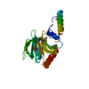
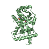
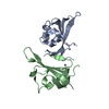
| Title | Crystal Structure of 30S Ribosomal Protein S8 from Aquifex Aeolicus | ||||||
 Components Components | 30S ribosomal protein S8 | ||||||
 Keywords Keywords | RIBOSOMAL PROTEIN / RNA-binding protein / Aquifex aeolicus / hyperthermophilic bacterium / Ribosome | ||||||
| Function / homology |  Function and homology information Function and homology informationcytosolic small ribosomal subunit / rRNA binding / structural constituent of ribosome / translation Similarity search - Function | ||||||
| Biological species |   Aquifex aeolicus (bacteria) Aquifex aeolicus (bacteria) | ||||||
| Method |  X-RAY DIFFRACTION / SYNCHROTRON / MAD, MOLECULAR REPLACEMENT / MAD / molecular replacement / Resolution: 2.16 Å X-RAY DIFFRACTION / SYNCHROTRON / MAD, MOLECULAR REPLACEMENT / MAD / molecular replacement / Resolution: 2.16 Å | ||||||
 Authors Authors | Menichelli, E. / Williamson, J.R. | ||||||
 Citation Citation | Journal: J.Mol.Biol. / Year: 2012 Title: The Structure of Aquifex aeolicus Ribosomal Protein S8 Reveals a Unique Subdomain that Contributes to an Extremely Tight Association with 16S rRNA. Authors: Menichelli, E. / Edgcomb, S.P. / Recht, M.I. / Williamson, J.R. | ||||||
| History |
|
- Structure visualization
| Structure viewer | Molecule: MolmilJmol/JSmol |
|---|
- Downloads & links
-Download
| PDBx/mmCIF format | 3rf2.cif.gz | 83.5 KB | Display | PDBx/mmCIF format |
|---|---|---|---|---|
| PDB format | pdb3rf2.ent.gz | 63.1 KB | Display | PDB format |
| PDBx/mmJSON format | 3rf2.json.gz | Tree view | PDBx/mmJSON format | |
| Others |  Other downloads Other downloads |
-Validation report
| Arichive directory | https://data.pdbj.org/pub/pdb/validation_reports/rf/3rf2ftp://data.pdbj.org/pub/pdb/validation_reports/rf/3rf2 | HTTPS FTP |
|---|
-Related structure data
| Similar structure data |
|---|


-Links
 PDBj
PDBj
- Assembly
| Deposited unit | 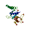
| ||||||||
|---|---|---|---|---|---|---|---|---|---|
| 1 |
| ||||||||
| Unit cell |
| ||||||||
| Components on special symmetry positions |
| ||||||||
| Details | AUTHORS STATE THAT THE PROTEIN WAS CHARACTERIZED BY SIZE EXCLUSION CHROMATOGRAPHY AND IN SOLUTION IT IS FOUND TO BE A MONOMER. |
-Components
| #1: Protein | Mass: 19494.951 Da / Num. of mol.: 1 Source method: isolated from a genetically manipulated source Source: (gene. exp.) Aquifex aeolicus (bacteria) / Gene: rpsH, aq_1651 / Plasmid: pET11c / Production host: | ||
|---|---|---|---|
| #2: Chemical | ChemComp-SO4 /   Mass: 96.063 Da / Num. of mol.: 5 / Source method: obtained synthetically / Formula: SO4 Mass: 96.063 Da / Num. of mol.: 5 / Source method: obtained synthetically / Formula: SO4#3: Water | ChemComp-HOH / |  Mass: 18.015 Da / Num. of mol.: 51 / Source method: isolated from a natural source / Formula: H2O Mass: 18.015 Da / Num. of mol.: 51 / Source method: isolated from a natural source / Formula: H2O |
-Experimental details
-Experiment
| Experiment | Method: X-RAY DIFFRACTION / Number of used crystals: 2 |
|---|
- Sample preparation
| Crystal | Density Matthews: 2.23 Å3/Da / Density % sol: 44.93 % |
|---|---|
| Crystal grow | Temperature: 295 K / Method: vapor diffusion / pH: 4.6 Details: 40% PEG 400, 0.1M sodium acetate, 0.2M lithium sulphate, pH 4.6, VAPOR DIFFUSION, temperature 295K |
-Data collection
| Diffraction |
| |||||||||||||||||||||||||||||||||||||||||||||||||||||||||||||||||||||||||||||
|---|---|---|---|---|---|---|---|---|---|---|---|---|---|---|---|---|---|---|---|---|---|---|---|---|---|---|---|---|---|---|---|---|---|---|---|---|---|---|---|---|---|---|---|---|---|---|---|---|---|---|---|---|---|---|---|---|---|---|---|---|---|---|---|---|---|---|---|---|---|---|---|---|---|---|---|---|---|---|
| Diffraction source |
| |||||||||||||||||||||||||||||||||||||||||||||||||||||||||||||||||||||||||||||
| Detector |
| |||||||||||||||||||||||||||||||||||||||||||||||||||||||||||||||||||||||||||||
| Radiation |
| |||||||||||||||||||||||||||||||||||||||||||||||||||||||||||||||||||||||||||||
| Radiation wavelength |
| |||||||||||||||||||||||||||||||||||||||||||||||||||||||||||||||||||||||||||||
| Reflection | Redundancy: 18.7 % / Av σ(I) over netI: 38.51 / Number: 208216 / Rmerge(I) obs: 0.122 / Χ2: 1.07 / D res high: 2.6 Å / D res low: 50 Å / Num. obs: 11135 / % possible obs: 98.6 | |||||||||||||||||||||||||||||||||||||||||||||||||||||||||||||||||||||||||||||
| Diffraction reflection shell |
| |||||||||||||||||||||||||||||||||||||||||||||||||||||||||||||||||||||||||||||
| Reflection | Resolution: 2.16→50 Å / Num. obs: 9918 / % possible obs: 99.3 % / Observed criterion σ(F): 0 / Observed criterion σ(I): 3 / Redundancy: 12.3 % / Rmerge(I) obs: 0.062 / Χ2: 0.994 / Net I/σ(I): 14.4 | |||||||||||||||||||||||||||||||||||||||||||||||||||||||||||||||||||||||||||||
| Reflection shell |
|

-Phasing
| Phasing |
| ||||||||||||||||||||||||||||||||||||||||||||||||||||||||
|---|---|---|---|---|---|---|---|---|---|---|---|---|---|---|---|---|---|---|---|---|---|---|---|---|---|---|---|---|---|---|---|---|---|---|---|---|---|---|---|---|---|---|---|---|---|---|---|---|---|---|---|---|---|---|---|---|---|
| Phasing MAD | D res high: 3 Å / D res low: 1000 Å / FOM : 0.62 / Reflection: 3907 | ||||||||||||||||||||||||||||||||||||||||||||||||||||||||
| Phasing MAD set site |
| ||||||||||||||||||||||||||||||||||||||||||||||||||||||||
| Phasing MAD shell |
| ||||||||||||||||||||||||||||||||||||||||||||||||||||||||
| Phasing MR |
| ||||||||||||||||||||||||||||||||||||||||||||||||||||||||
| Phasing dm | FOM : 0.72 / FOM acentric: 0.73 / FOM centric: 0.69 / Reflection: 3900 / Reflection acentric: 2980 / Reflection centric: 920 | ||||||||||||||||||||||||||||||||||||||||||||||||||||||||
| Phasing dm shell |
|
- Processing
| Software |
| |||||||||||||||||||||||||||||||||||||||||||||||||||||||||||||||||||||||||||||||||||||||||||||||||||||||||||||||||||||||||||||||||||||||||||||||||||||||||||||||||||||||||||||||
|---|---|---|---|---|---|---|---|---|---|---|---|---|---|---|---|---|---|---|---|---|---|---|---|---|---|---|---|---|---|---|---|---|---|---|---|---|---|---|---|---|---|---|---|---|---|---|---|---|---|---|---|---|---|---|---|---|---|---|---|---|---|---|---|---|---|---|---|---|---|---|---|---|---|---|---|---|---|---|---|---|---|---|---|---|---|---|---|---|---|---|---|---|---|---|---|---|---|---|---|---|---|---|---|---|---|---|---|---|---|---|---|---|---|---|---|---|---|---|---|---|---|---|---|---|---|---|---|---|---|---|---|---|---|---|---|---|---|---|---|---|---|---|---|---|---|---|---|---|---|---|---|---|---|---|---|---|---|---|---|---|---|---|---|---|---|---|---|---|---|---|---|---|---|---|---|---|
| Refinement | Method to determine structure: MAD, MOLECULAR REPLACEMENT / Resolution: 2.16→30.432 Å / Occupancy max: 1 / Occupancy min: 0 / FOM work R set: 0.7713 / SU ML: 0.31 / σ(F): 0 / Stereochemistry target values: ML
| |||||||||||||||||||||||||||||||||||||||||||||||||||||||||||||||||||||||||||||||||||||||||||||||||||||||||||||||||||||||||||||||||||||||||||||||||||||||||||||||||||||||||||||||
| Solvent computation | Shrinkage radii: 0.83 Å / VDW probe radii: 1.1 Å / Solvent model: FLAT BULK SOLVENT MODEL / Bsol: 60.458 Å2 / ksol: 0.361 e/Å3 | |||||||||||||||||||||||||||||||||||||||||||||||||||||||||||||||||||||||||||||||||||||||||||||||||||||||||||||||||||||||||||||||||||||||||||||||||||||||||||||||||||||||||||||||
| Displacement parameters | Biso max: 170.6 Å2 / Biso mean: 51.4623 Å2 / Biso min: 16.08 Å2
| |||||||||||||||||||||||||||||||||||||||||||||||||||||||||||||||||||||||||||||||||||||||||||||||||||||||||||||||||||||||||||||||||||||||||||||||||||||||||||||||||||||||||||||||
| Refinement step | Cycle: LAST / Resolution: 2.16→30.432 Å
| |||||||||||||||||||||||||||||||||||||||||||||||||||||||||||||||||||||||||||||||||||||||||||||||||||||||||||||||||||||||||||||||||||||||||||||||||||||||||||||||||||||||||||||||
| Refine LS restraints |
| |||||||||||||||||||||||||||||||||||||||||||||||||||||||||||||||||||||||||||||||||||||||||||||||||||||||||||||||||||||||||||||||||||||||||||||||||||||||||||||||||||||||||||||||
| LS refinement shell | Refine-ID: X-RAY DIFFRACTION / Total num. of bins used: 7
| |||||||||||||||||||||||||||||||||||||||||||||||||||||||||||||||||||||||||||||||||||||||||||||||||||||||||||||||||||||||||||||||||||||||||||||||||||||||||||||||||||||||||||||||
| Refinement TLS params. | Method: refined / Refine-ID: X-RAY DIFFRACTION
| |||||||||||||||||||||||||||||||||||||||||||||||||||||||||||||||||||||||||||||||||||||||||||||||||||||||||||||||||||||||||||||||||||||||||||||||||||||||||||||||||||||||||||||||
| Refinement TLS group |
|
