 Movie
Movie Controller


+ Open data

- Basic information
| Entry | Database: PDB / ID: 3n1j | ||||||
|---|---|---|---|---|---|---|---|
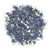
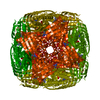
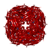
| Title | Crystal structure of a StWhy2-dT32 complex | ||||||
 Components Components |
| ||||||
 Keywords Keywords | DNA BINDING PROTEIN/DNA / Single-stranded DNA binding protein / Plant / Whirly / Protein-DNA complex / DNA BINDING PROTEIN-DNA complex | ||||||
| Function / homology |  Function and homology information Function and homology informationdefense response / single-stranded DNA binding / DNA repair / regulation of DNA-templated transcription / mitochondrion / DNA binding Similarity search - Function | ||||||
| Biological species |  | ||||||
| Method |  X-RAY DIFFRACTION / SYNCHROTRON / MOLECULAR REPLACEMENT / Resolution: 2.65 Å X-RAY DIFFRACTION / SYNCHROTRON / MOLECULAR REPLACEMENT / Resolution: 2.65 Å | ||||||
 Authors Authors | Cappadocia, L. / Brisson, N. / Sygusch, J. | ||||||
 Citation Citation | Journal: Plant Cell / Year: 2010 Title: Crystal Structures of DNA-Whirly Complexes and Their Role in Arabidopsis Organelle Genome Repair. Authors: Cappadocia, L. / Marechal, A. / Parent, J.S. / Lepage, E. / Sygusch, J. / Brisson, N. | ||||||
| History |
|
- Structure visualization
| Structure viewer | Molecule: MolmilJmol/JSmol |
|---|
- Downloads & links
-Download
| PDBx/mmCIF format | 3n1j.cif.gz | 52.3 KB | Display | PDBx/mmCIF format |
|---|---|---|---|---|
| PDB format | pdb3n1j.ent.gz | 38.2 KB | Display | PDB format |
| PDBx/mmJSON format | 3n1j.json.gz | Tree view | PDBx/mmJSON format | |
| Others |  Other downloads Other downloads |
-Validation report
| Arichive directory | https://data.pdbj.org/pub/pdb/validation_reports/n1/3n1jftp://data.pdbj.org/pub/pdb/validation_reports/n1/3n1j | HTTPS FTP |
|---|
-Related structure data
| Related structure data | 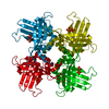 3n1hC 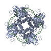 3n1iC  3n1kC 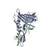 3n1lC  1l3aS C: citing same article ( S: Starting model for refinement |
|---|---|
| Similar structure data |

-Links
 PDBj
PDBj- Assembly
| Deposited unit | 
| ||||||||
|---|---|---|---|---|---|---|---|---|---|
| 1 | x 24
| ||||||||
| 2 | 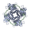
| ||||||||
| Unit cell |
| ||||||||
| Details | Authors state that the biological unit is a tetramer protein plus a 32-mer DNA molecule, as indicated as pentamer in remark 350. Please refer to remark 999 for more details on the sequences specificity of this crystal structure. |
-Components
| #1: Protein | Mass: 20000.715 Da / Num. of mol.: 1 Source method: isolated from a genetically manipulated source Source: (gene. exp.)  |
|---|---|
| #2: DNA chain | Mass: 2692.778 Da / Num. of mol.: 1 / Source method: obtained synthetically / Details: DNA Synthesis |
| #3: Water | ChemComp-HOH /  Mass: 18.015 Da / Num. of mol.: 60 / Source method: isolated from a natural source / Formula: H2O Mass: 18.015 Da / Num. of mol.: 60 / Source method: isolated from a natural source / Formula: H2O |
| Sequence details | THE SEQUENCE OF THE CRYSTALLIZED DNA IS A 32-MER OLIGONUCLEOTIDE WITH SEQUENCE ...THE SEQUENCE OF THE CRYSTALLIZ |
-Experimental details
-Experiment
| Experiment | Method: X-RAY DIFFRACTION / Number of used crystals: 1 |
|---|
- Sample preparation
| Crystal | Density Matthews: 2.14 Å3/Da / Density % sol: 47.29 % |
|---|---|
| Crystal grow | Temperature: 298 K / Method: vapor diffusion, hanging drop / pH: 8 Details: 25% PEG6000, 0.1M Tris, 1.2M LiCl, pH 8.0, VAPOR DIFFUSION, HANGING DROP, temperature 298K |
-Data collection
| Diffraction | Mean temperature: 100 K |
|---|---|
| Diffraction source | Source: SYNCHROTRON / Site: NSLS  / Beamline: X25 / Wavelength: 1.08 Å / Beamline: X25 / Wavelength: 1.08 Å |
| Detector | Type: ADSC QUANTUM 315 / Detector: CCD / Date: Nov 13, 2007 |
| Radiation | Protocol: SINGLE WAVELENGTH / Monochromatic (M) / Laue (L): M / Scattering type: x-ray |
| Radiation wavelength | Wavelength: 1.08 Å / Relative weight: 1 |
| Reflection | Resolution: 2.65→50 Å / Num. all: 6196 / Num. obs: 6188 / % possible obs: 100 % / Observed criterion σ(F): 0 / Observed criterion σ(I): 0 / Redundancy: 19.1 % / Biso Wilson estimate: 52 Å2 / Rsym value: 0.118 / Net I/σ(I): 9.1 |
| Reflection shell | Resolution: 2.65→2.74 Å / Redundancy: 12.7 % / Mean I/σ(I) obs: 3 / Num. unique all: 605 / Rsym value: 0.87 / % possible all: 100 |
- Processing
| Software |
| |||||||||||||||||||||||||||||||||||
|---|---|---|---|---|---|---|---|---|---|---|---|---|---|---|---|---|---|---|---|---|---|---|---|---|---|---|---|---|---|---|---|---|---|---|---|---|
| Refinement | Method to determine structure: MOLECULAR REPLACEMENT Starting model: PDB ENTRY 1L3A Resolution: 2.65→38.197 Å / SU ML: 2.53 / Isotropic thermal model: Isotropic / Cross valid method: THROUGHOUT / σ(F): 0.05 / Stereochemistry target values: ML
| |||||||||||||||||||||||||||||||||||
| Solvent computation | Shrinkage radii: 0.9 Å / VDW probe radii: 1.11 Å / Solvent model: FLAT BULK SOLVENT MODEL / Bsol: 48.914 Å2 / ksol: 0.307 e/Å3 | |||||||||||||||||||||||||||||||||||
| Displacement parameters | Biso mean: 54.37 Å2
| |||||||||||||||||||||||||||||||||||
| Refinement step | Cycle: LAST / Resolution: 2.65→38.197 Å
| |||||||||||||||||||||||||||||||||||
| Refine LS restraints |
| |||||||||||||||||||||||||||||||||||
| LS refinement shell |
|
