- PDB-3ra0: Crystal Structure of a StWhy2 K67A-dT32 complex -
+
Open data
ID or keywords:
Loading...
-
Basic information
Entry
Database: PDB / ID: 3ra0
Title
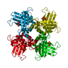
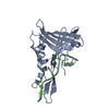
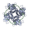
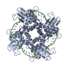
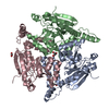
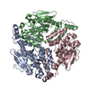
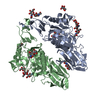
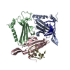
Crystal Structure of a StWhy2 K67A-dT32 complex
Components
DNA 32-mer dT32
Why2 protein
Keywords
DNA BINDING PROTEIN/DNA / StWhy2 / single-stranded DNA binding protein / Plant / Potato / Whirly / protein-DNA complex / Mitochondria / DNA BINDING PROTEIN-DNA complex
Function / homology
Function and homology information
defense response / single-stranded DNA binding / DNA repair / regulation of DNA-templated transcription / mitochondrion / DNA binding Similarity search - Function
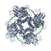
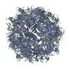
Authors state that the biological unit is a tetramer protein plus a 32-mer DNA molecule, as indicated as pentamer in remark 350. Please refer to remark 999 for more details on the sequences specificity of this crystal structure.
-
Components
#1: Protein
Why2protein
Mass: 19942.613 Da / Num. of mol.: 1 / Fragment: Residues 48-216 / Mutation: K67A Source method: isolated from a genetically manipulated source Source: (gene. exp.) Solanum tuberosum (potato) / Strain: kennebec / Gene: StWhy2 / Plasmid: pET21a / Production host: Escherichia coli (E. coli) / Strain (production host): BL21(DE3) / References: UniProt: D9J034
#2: DNA chain
DNA32-merdT32
Mass: 9689.213 Da / Num. of mol.: 1 / Source method: obtained synthetically / Details: DNA synthesis
Mass: 18.015 Da / Num. of mol.: 30 / Source method: isolated from a natural source / Formula: H2O
Sequence details
THE SEQUENCE OF THE CRYSTALLIZED DNA IS A 32-MER OLIGONUCLEOTIDE WITH SEQUENCE ...THE SEQUENCE OF THE CRYSTALLIZED DNA IS A 32-MER OLIGONUCLEOTIDE WITH SEQUENCE TTTTTTTTTTTTTTTTTTTTTTTTTTTTTTTT. ONLY 9 RESIDUES WERE MODELED IN THE COORDINATES OF THE ASYMMETRIC UNIT. ACCORDING TO THE AUTHORS, THE 32-MER DNA BINDS A TETRAMER THAT INCLUDE FOUR MONOMERS OF FOUR ASYMMETRIC UNITS; THE STWHY2-DNA BINDING IS NOT SEQUENCE SPECIFIC THUS EACH TETRAMERIC PROTEIN BINDS A 32-MER DNA IN DIFFERENT SEQUENCE REGISTERS; AS A RESULT ONLY THREE OF THE FOUR DNA-BINDING SITES OF EACH TETRAMER WOULD BE PHYSICALLY OCCUPIED BY THE DNA.
-
Experimental details
-
Experiment
Experiment
Method: X-RAY DIFFRACTION / Number of used crystals: 1
-
Sample preparation
Crystal
Density Matthews: 2.14 Å3/Da / Density % sol: 47.29 %
Crystal grow
Temperature: 298 K / Method: vapor diffusion, hanging drop / pH: 8 Details: 100mM Tris pH8.0, 15% PEG6000, 1200mM LiCl, vapor diffusion, hanging drop, temperature 298K
In the structure databanks used in Yorodumi, some data are registered as the other names, "COVID-19 virus" and "2019-nCoV". Here are the details of the virus and the list of structure data.
Jan 31, 2019. EMDB accession codes are about to change! (news from PDBe EMDB page)
EMDB accession codes are about to change! (news from PDBe EMDB page)
The allocation of 4 digits for EMDB accession codes will soon come to an end. Whilst these codes will remain in use, new EMDB accession codes will include an additional digit and will expand incrementally as the available range of codes is exhausted. The current 4-digit format prefixed with “EMD-” (i.e. EMD-XXXX) will advance to a 5-digit format (i.e. EMD-XXXXX), and so on. It is currently estimated that the 4-digit codes will be depleted around Spring 2019, at which point the 5-digit format will come into force.
The EM Navigator/Yorodumi systems omit the EMD- prefix.
Related info.:Q: What is EMD? / ID/Accession-code notation in Yorodumi/EM Navigator
Yorodumi is a browser for structure data from EMDB, PDB, SASBDB, etc.
This page is also the successor to EM Navigator detail page, and also detail information page/front-end page for Omokage search.
The word "yorodu" (or yorozu) is an old Japanese word meaning "ten thousand". "mi" (miru) is to see.
Related info.:EMDB / PDB / SASBDB / Comparison of 3 databanks / Yorodumi Search / Aug 31, 2016. New EM Navigator & Yorodumi / Yorodumi Papers / Jmol/JSmol / Function and homology information / Changes in new EM Navigator and Yorodumi
 Movie
Movie Controller


 Open data
 Basic information
 Components
 Keywords
 Function and homology information
 X-RAY DIFFRACTION /
  Authors
 Citation
 Structure visualization
 Downloads & links
 Other downloads


 PDBj
PDBj


 Assembly
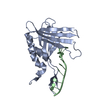

 Mass: 18.015 Da / Num. of mol.: 30 / Source method: isolated from a natural source / Formula: H2O
 Sample preparation
 / Beamline: X29A / Wavelength: 1.08 Å
 Processing