 Movie
Movie Controller

[English] 日本語
 Yorodumi
- PDB-2hqv: X-ray Crystal Structure of Protein AGR_C_4470 from Agrobacterium ... -
+ Open data

- Basic information
| Entry | Database: PDB / ID: 2hqv | ||||||
|---|---|---|---|---|---|---|---|
| Title | X-ray Crystal Structure of Protein AGR_C_4470 from Agrobacterium tumefaciens. Northeast Structural Genomics Consortium Target AtR92. | ||||||
 Components Components | AGR_C_4470p | ||||||
 Keywords Keywords | STRUCTURAL GENOMICS / UNKNOWN FUNCTION / NESG / AtR92 / AGR_C_4470 / Q7CX01_AGRT5 / PSI-2 / Protein Structure Initiative / Northeast Structural Genomics Consortium | ||||||
| Function / homology | Intracellular heme transport protein HutX-like / Haem utilisation ChuX/HutX / HemS/ChuS/ChuX like domains / Heme iron utilization protein-like fold / : / 3-Layer(aba) Sandwich / Alpha Beta / Heme utilization cystosolic carrier protein HutX Function and homology information Function and homology information | ||||||
| Biological species |  Agrobacterium tumefaciens str. C58 (bacteria) Agrobacterium tumefaciens str. C58 (bacteria) | ||||||
| Method |  X-RAY DIFFRACTION / SYNCHROTRON / MAD / Resolution: 2 Å X-RAY DIFFRACTION / SYNCHROTRON / MAD / Resolution: 2 Å | ||||||
 Authors Authors | Vorobiev, S.M. / Neely, H. / Seetharaman, J. / Zhao, L. / Cunningham, K. / Ma, L.C. / Fang, Y. / Xiao, R. / Acton, T. / Montelione, T.G. ...Vorobiev, S.M. / Neely, H. / Seetharaman, J. / Zhao, L. / Cunningham, K. / Ma, L.C. / Fang, Y. / Xiao, R. / Acton, T. / Montelione, T.G. / Hunt, J.F. / Tong, L. / Northeast Structural Genomics Consortium (NESG) | ||||||
 Citation Citation | Journal: Protein Sci. / Year: 2007 Title: Crystal structure of AGR_C_4470p from Agrobacterium tumefaciens. Authors: Vorobiev, S.M. / Neely, H. / Seetharaman, J. / Ma, L.C. / Xiao, R. / Acton, T.B. / Montelione, G.T. / Tong, L. | ||||||
| History |
|
- Structure visualization
| Structure viewer | Molecule: MolmilJmol/JSmol |
|---|
- Downloads & links
-Download
| PDBx/mmCIF format | 2hqv.cif.gz | 46.7 KB | Display | PDBx/mmCIF format |
|---|---|---|---|---|
| PDB format | pdb2hqv.ent.gz | 32.3 KB | Display | PDB format |
| PDBx/mmJSON format | 2hqv.json.gz | Tree view | PDBx/mmJSON format | |
| Others |  Other downloads Other downloads |
-Validation report
| Arichive directory | https://data.pdbj.org/pub/pdb/validation_reports/hq/2hqvftp://data.pdbj.org/pub/pdb/validation_reports/hq/2hqv | HTTPS FTP |
|---|
-Related structure data
| Similar structure data | |
|---|---|
| Other databases |


-Links
 PDBj
PDBj- Assembly
| Deposited unit | 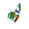
| ||||||||
|---|---|---|---|---|---|---|---|---|---|
| 1 | 
| ||||||||
| Unit cell |
| ||||||||
| Details | Dimer. The second part of the dimer is generated by -X, -Y, Z + (1 0 0) operator. |
-Components
| #1: Protein | Mass: 21620.748 Da / Num. of mol.: 1 / Mutation: C-tag: LEHHHHHH Source method: isolated from a genetically manipulated source Source: (gene. exp.) Agrobacterium tumefaciens str. C58 (bacteria)Species: Agrobacterium tumefaciens / Strain: C58-ATCC 33970 / Gene: AGR_C_4470 / Plasmid: pET21 / Production host: |
|---|---|
| #2: Water | ChemComp-HOH /  Mass: 18.015 Da / Num. of mol.: 67 / Source method: isolated from a natural source / Formula: H2O Mass: 18.015 Da / Num. of mol.: 67 / Source method: isolated from a natural source / Formula: H2O |
| Has protein modification | Y |
-Experimental details
-Experiment
| Experiment | Method: X-RAY DIFFRACTION / Number of used crystals: 1 |
|---|
- Sample preparation
| Crystal | Density Matthews: 2.9 Å3/Da / Density % sol: 57.57 % Description: THE STRUCTURE FACTOR FILE CONTAINS FRIEDEL PAIRS |
|---|---|
| Crystal grow | Temperature: 291 K / Method: vapor diffusion, hanging drop / pH: 6 Details: 40% PEG 400, 0.1M Magnesium sulfate, 0.1M MES, pH 6.0, VAPOR DIFFUSION, HANGING DROP, temperature 291K |
-Data collection
| Diffraction | Mean temperature: 100 K | |||||||||||||||
|---|---|---|---|---|---|---|---|---|---|---|---|---|---|---|---|---|
| Diffraction source | Source: SYNCHROTRON / Site: NSLS  / Beamline: X4A / Wavelength: 0.97960, 0.97923, 0.97947, 0.96783 / Beamline: X4A / Wavelength: 0.97960, 0.97923, 0.97947, 0.96783 | |||||||||||||||
| Detector | Type: ADSC QUANTUM 4 / Detector: CCD / Date: Jul 13, 2006 | |||||||||||||||
| Radiation | Protocol: MAD / Monochromatic (M) / Laue (L): M / Scattering type: x-ray | |||||||||||||||
| Radiation wavelength |
| |||||||||||||||
| Reflection | Resolution: 2→30 Å / Num. all: 32944 / Num. obs: 32911 / % possible obs: 99.9 % / Observed criterion σ(F): 0 / Observed criterion σ(I): 0 / Redundancy: 7.5 % / Biso Wilson estimate: 21.3 Å2 / Rmerge(I) obs: 0.067 / Net I/σ(I): 28.5 | |||||||||||||||
| Reflection shell | Resolution: 2→2.07 Å / Redundancy: 6.5 % / Rmerge(I) obs: 0.544 / Mean I/σ(I) obs: 3.4 / % possible all: 99.9 |
- Processing
| Software |
| ||||||||||||||||||||||||||||||||||||||||||||||||||||||||||||
|---|---|---|---|---|---|---|---|---|---|---|---|---|---|---|---|---|---|---|---|---|---|---|---|---|---|---|---|---|---|---|---|---|---|---|---|---|---|---|---|---|---|---|---|---|---|---|---|---|---|---|---|---|---|---|---|---|---|---|---|---|---|
| Refinement | Method to determine structure: MAD / Resolution: 2→29.76 Å / Rfactor Rfree error: 0.008 / Data cutoff high absF: 109456.86 / Data cutoff low absF: 0 / Isotropic thermal model: RESTRAINED / Cross valid method: THROUGHOUT / σ(F): 2 / Stereochemistry target values: Engh & Huber / Details: THE FRIEDEL PAIRS WERE USED FOR REFINEMENT
| ||||||||||||||||||||||||||||||||||||||||||||||||||||||||||||
| Solvent computation | Solvent model: FLAT MODEL / Bsol: 55.1963 Å2 / ksol: 0.368725 e/Å3 | ||||||||||||||||||||||||||||||||||||||||||||||||||||||||||||
| Displacement parameters | Biso mean: 42 Å2
| ||||||||||||||||||||||||||||||||||||||||||||||||||||||||||||
| Refine analyze |
| ||||||||||||||||||||||||||||||||||||||||||||||||||||||||||||
| Refinement step | Cycle: LAST / Resolution: 2→29.76 Å
| ||||||||||||||||||||||||||||||||||||||||||||||||||||||||||||
| Refine LS restraints |
| ||||||||||||||||||||||||||||||||||||||||||||||||||||||||||||
| LS refinement shell | Resolution: 2→2.13 Å / Rfactor Rfree error: 0.018 / Total num. of bins used: 6
| ||||||||||||||||||||||||||||||||||||||||||||||||||||||||||||
| Xplor file |
|
