 Movie
Movie Controller

[English] 日本語
 Yorodumi
- PDB-3r9i: 2.6A resolution structure of MinD complexed with MinE (12-31) peptide -
+ Open data

- Basic information
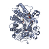
| Entry | Database: PDB / ID: 3r9i | ||||||
|---|---|---|---|---|---|---|---|
| Title | 2.6A resolution structure of MinD complexed with MinE (12-31) peptide | ||||||
 Components Components |
| ||||||
 Keywords Keywords | CELL CYCLE / HYDROLASE/CELL CYCLE / ATPase / bacterial cell division inhibitor / mine / HYDROLASE-CELL CYCLE complex | ||||||
| Function / homology |  Function and homology information Function and homology informationregulation of division septum assembly / division septum site selection / cell pole / intracellular mRNA localization / ATPase activator activity / chromosome segregation / cytoplasmic side of plasma membrane / cell division / ATP hydrolysis activity / ATP binding ...regulation of division septum assembly / division septum site selection / cell pole / intracellular mRNA localization / ATPase activator activity / chromosome segregation / cytoplasmic side of plasma membrane / cell division / ATP hydrolysis activity / ATP binding / identical protein binding / plasma membrane / cytosol Similarity search - Function | ||||||
| Biological species |  | ||||||
| Method |  X-RAY DIFFRACTION / SYNCHROTRON / MOLECULAR REPLACEMENT / molecular replacement / Resolution: 2.6 Å X-RAY DIFFRACTION / SYNCHROTRON / MOLECULAR REPLACEMENT / molecular replacement / Resolution: 2.6 Å | ||||||
 Authors Authors | Lovell, S. / Battaile, K.P. / Park, K.-T. / Wu, W. / Holyoak, T. / Lutkenhaus, J. | ||||||
 Citation Citation | Journal: Cell(Cambridge,Mass.) / Year: 2011 Title: The Min Oscillator Uses MinD-Dependent Conformational Changes in MinE to Spatially Regulate Cytokinesis. Authors: Park, K.T. / Wu, W. / Battaile, K.P. / Lovell, S. / Holyoak, T. / Lutkenhaus, J. | ||||||
| History |
|
- Structure visualization
| Structure viewer | Molecule: MolmilJmol/JSmol |
|---|
- Downloads & links
-Download
| PDBx/mmCIF format | 3r9i.cif.gz | 216.9 KB | Display | PDBx/mmCIF format |
|---|---|---|---|---|
| PDB format | pdb3r9i.ent.gz | 173.6 KB | Display | PDB format |
| PDBx/mmJSON format | 3r9i.json.gz | Tree view | PDBx/mmJSON format | |
| Others |  Other downloads Other downloads |
-Validation report
| Arichive directory | https://data.pdbj.org/pub/pdb/validation_reports/r9/3r9iftp://data.pdbj.org/pub/pdb/validation_reports/r9/3r9i | HTTPS FTP |
|---|
-Related structure data
| Related structure data | 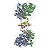 3r9jC 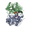 3q9lS S: Starting model for refinement C: citing same article ( |
|---|---|
| Similar structure data |


-Links
 PDBj
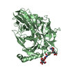
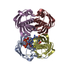
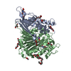
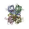
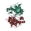
PDBj- Assembly
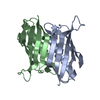
| Deposited unit | 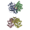
| |||||||||||||||||||||||||||||||||||||||||||||||||||||||||||||||||||||||||||||||
|---|---|---|---|---|---|---|---|---|---|---|---|---|---|---|---|---|---|---|---|---|---|---|---|---|---|---|---|---|---|---|---|---|---|---|---|---|---|---|---|---|---|---|---|---|---|---|---|---|---|---|---|---|---|---|---|---|---|---|---|---|---|---|---|---|---|---|---|---|---|---|---|---|---|---|---|---|---|---|---|---|
| 1 | 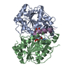
| |||||||||||||||||||||||||||||||||||||||||||||||||||||||||||||||||||||||||||||||
| 2 | 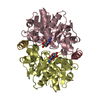
| |||||||||||||||||||||||||||||||||||||||||||||||||||||||||||||||||||||||||||||||
| Unit cell |
| |||||||||||||||||||||||||||||||||||||||||||||||||||||||||||||||||||||||||||||||
| Noncrystallographic symmetry (NCS) | NCS domain:
NCS domain segments:
|