 Movie
Movie Controller


+ Open data

- Basic information
| Entry | Database: PDB / ID: 1n9m | ||||||
|---|---|---|---|---|---|---|---|
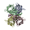
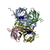
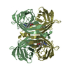
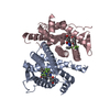
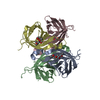
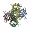
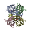
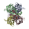
| Title | Streptavidin Mutant S27A with Biotin at 1.6A Resolution | ||||||
 Components Components | Streptavidin | ||||||
 Keywords Keywords | BIOTIN-BINDING PROTEIN / homotetramer | ||||||
| Function / homology |  Function and homology information Function and homology information | ||||||
| Biological species |  Streptomyces avidinii (bacteria) Streptomyces avidinii (bacteria) | ||||||
| Method |  X-RAY DIFFRACTION / isomorphous / Resolution: 1.6 Å X-RAY DIFFRACTION / isomorphous / Resolution: 1.6 Å | ||||||
 Authors Authors | Le Trong, I. / Freitag, S. / Klumb, L.A. / Chu, V. / Stayton, P.S. / Stenkamp, R.E. | ||||||
 Citation Citation | Journal: Acta Crystallogr.,Sect.D / Year: 2003 Title: Structural studies of hydrogen bonds in the high-affinity streptavidin-biotin complex: mutations of amino acids interacting with the ureido oxygen of biotin. Authors: Le Trong, I. / Freitag, S. / Klumb, L.A. / Chu, V. / Stayton, P.S. / Stenkamp, R.E. | ||||||
| History |
|
- Structure visualization
| Structure viewer | Molecule: MolmilJmol/JSmol |
|---|
- Downloads & links
-Download
| PDBx/mmCIF format | 1n9m.cif.gz | 211 KB | Display | PDBx/mmCIF format |
|---|---|---|---|---|
| PDB format | pdb1n9m.ent.gz | 170 KB | Display | PDB format |
| PDBx/mmJSON format | 1n9m.json.gz | Tree view | PDBx/mmJSON format | |
| Others |  Other downloads Other downloads |
-Validation report
| Arichive directory | https://data.pdbj.org/pub/pdb/validation_reports/n9/1n9mftp://data.pdbj.org/pub/pdb/validation_reports/n9/1n9m | HTTPS FTP |
|---|
-Related structure data
| Related structure data |  1n43C  1n4jC  1n7yC 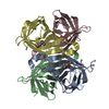 1n9yC 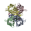 1nbxC 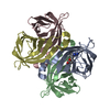 1nc9C 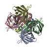 1ndjC C: citing same article ( |
|---|---|
| Similar structure data |


-Links
 PDBj
PDBj- Assembly
| Deposited unit | 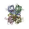
| ||||||||
|---|---|---|---|---|---|---|---|---|---|
| 1 |
| ||||||||
| Unit cell |
|
-Components
| #1: Protein | Mass: 13265.336 Da / Num. of mol.: 4 / Fragment: core streptavidin, residues 13-139 / Mutation: S27A Source method: isolated from a genetically manipulated source Source: (gene. exp.) Streptomyces avidinii (bacteria) / Gene: core streptavidin / Plasmid: pET21a / Species (production host): Escherichia coli / Production host: #2: Chemical | ChemComp-BTN /   Mass: 244.311 Da / Num. of mol.: 4 / Source method: obtained synthetically / Formula: C10H16N2O3S Mass: 244.311 Da / Num. of mol.: 4 / Source method: obtained synthetically / Formula: C10H16N2O3S#3: Water | ChemComp-HOH / |  Mass: 18.015 Da / Num. of mol.: 459 / Source method: isolated from a natural source / Formula: H2O Mass: 18.015 Da / Num. of mol.: 459 / Source method: isolated from a natural source / Formula: H2O |
|---|
-Experimental details
-Experiment
| Experiment | Method: X-RAY DIFFRACTION / Number of used crystals: 1 |
|---|
- Sample preparation
| Crystal | Density Matthews: 2.45 Å3/Da / Density % sol: 52.69 % | |||||||||||||||
|---|---|---|---|---|---|---|---|---|---|---|---|---|---|---|---|---|
| Crystal grow | Temperature: 293 K / Method: vapor diffusion, sitting drop / pH: 4.5 Details: MPD, pH 4.5, VAPOR DIFFUSION, SITTING DROP, temperature 293K | |||||||||||||||
| Crystal grow | *PLUS Method: vapor diffusion, hanging drop | |||||||||||||||
| Components of the solutions | *PLUS
|
-Data collection
| Diffraction | Mean temperature: 120 K |
|---|---|
| Diffraction source | Source: ROTATING ANODE / Type: RIGAKU RU200 / Wavelength: 1.5418 Å |
| Detector | Type: RIGAKU RAXIS II / Detector: IMAGE PLATE / Date: Jul 1, 1998 / Details: mirrors |
| Radiation | Monochromator: mirrors / Protocol: SINGLE WAVELENGTH / Monochromatic (M) / Laue (L): M / Scattering type: x-ray |
| Radiation wavelength | Wavelength: 1.5418 Å / Relative weight: 1 |
| Reflection | Resolution: 1.6→50 Å / Num. all: 55581 / Num. obs: 55581 / % possible obs: 68.5 % / Observed criterion σ(F): 0 / Observed criterion σ(I): 0 / Rmerge(I) obs: 0.041 |
| Reflection | *PLUS Highest resolution: 1.6 Å / % possible obs: 68 % / Rmerge(I) obs: 0.04 |
| Reflection shell | *PLUS % possible obs: 0.1 % |
- Processing
| Software |
| |||||||||||||||||||||||||||||||||
|---|---|---|---|---|---|---|---|---|---|---|---|---|---|---|---|---|---|---|---|---|---|---|---|---|---|---|---|---|---|---|---|---|---|---|
| Refinement | Method to determine structure: isomorphous / Resolution: 1.6→10 Å / Num. parameters: 37440 / Num. restraintsaints: 46215 / Cross valid method: FREE R / σ(F): 0 / Stereochemistry target values: ENGH AND HUBER Details: ANISOTROPIC REFINEMENT REDUCED FREE R (NO CUTOFF) BY ?
| |||||||||||||||||||||||||||||||||
| Refine analyze | Num. disordered residues: 4 / Occupancy sum hydrogen: 0 / Occupancy sum non hydrogen: 4156 | |||||||||||||||||||||||||||||||||
| Refinement step | Cycle: LAST / Resolution: 1.6→10 Å
| |||||||||||||||||||||||||||||||||
| Refine LS restraints |
| |||||||||||||||||||||||||||||||||
| Software | *PLUS Name: SHELXL / Version: 97 / Classification: refinement | |||||||||||||||||||||||||||||||||
| Refinement | *PLUS Highest resolution: 1.6 Å / Lowest resolution: 10 Å / Rfactor Rfree: 0.248 / Rfactor Rwork: 0.159 / % reflection Rfree: 10 % | |||||||||||||||||||||||||||||||||
| Solvent computation | *PLUS | |||||||||||||||||||||||||||||||||
| Displacement parameters | *PLUS |
