 Movie
Movie Controller

[English] 日本語
 Yorodumi
- PDB-3qob: Mechanical Coupling Controls Cooperative Ligand Binding in a Homo... -
+ Open data

- Basic information
| Entry | Database: PDB / ID: 3qob | ||||||
|---|---|---|---|---|---|---|---|
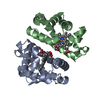
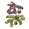
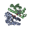
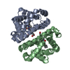
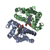
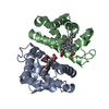
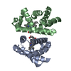
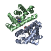
| Title | Mechanical Coupling Controls Cooperative Ligand Binding in a Homodimeric Hemoglobin | ||||||
 Components Components | Globin-1 | ||||||
 Keywords Keywords | OXYGEN TRANSPORT / time-resolved / Laue diffraction / dynamic crystallography / heterogeneous structure | ||||||
| Function / homology |  Function and homology information Function and homology informationoxygen carrier activity / oxygen binding / heme binding / metal ion binding / identical protein binding / cytoplasm Similarity search - Function | ||||||
| Biological species |  Anadara inaequivalvis (ark clam) Anadara inaequivalvis (ark clam) | ||||||
| Method |  X-RAY DIFFRACTION / SYNCHROTRON / FOURIER SYNTHESIS / Resolution: 1.6 Å X-RAY DIFFRACTION / SYNCHROTRON / FOURIER SYNTHESIS / Resolution: 1.6 Å | ||||||
 Authors Authors | Ren, Z. | ||||||
 Citation Citation | Journal: Proc.Natl.Acad.Sci.USA / Year: 2012 Title: Cooperative macromolecular device revealed by meta-analysis of static and time-resolved structures. Authors: Ren, Z. / Srajer, V. / Knapp, J.E. / Royer Jr., W.E. | ||||||
| History |
|
- Structure visualization
| Structure viewer | Molecule: MolmilJmol/JSmol |
|---|
- Downloads & links
-Download
| PDBx/mmCIF format | 3qob.cif.gz | 62.3 KB | Display | PDBx/mmCIF format |
|---|---|---|---|---|
| PDB format | pdb3qob.ent.gz | 45 KB | Display | PDB format |
| PDBx/mmJSON format | 3qob.json.gz | Tree view | PDBx/mmJSON format | |
| Others |  Other downloads Other downloads |
-Validation report
| Arichive directory | https://data.pdbj.org/pub/pdb/validation_reports/qo/3qobftp://data.pdbj.org/pub/pdb/validation_reports/qo/3qob | HTTPS FTP |
|---|
-Related structure data
| Related structure data | 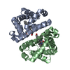 3ugyC  3ugzC  3uh3C  3uh5C  3uh6C  3uh7C  3uhbC  3uhcC  3uhdC  3uheC  3uhgC  3uhhC  3uhiC  3uhkC  3uhnC  3uhqC  3uhrC  3uhsC  3uhtC  3uhuC  3uhvC  3uhwC  3uhxC  3uhyC  3uhzC  3ui0C 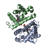 3sdhS S: Starting model for refinement C: citing same article ( |
|---|---|
| Similar structure data |


-Links
 PDBj
PDBj


- Assembly
| Deposited unit | 
| ||||||||
|---|---|---|---|---|---|---|---|---|---|
| 1 |
| ||||||||
| Unit cell |
|
-Components
| #1: Protein | Mass: 15966.319 Da / Num. of mol.: 2 / Source method: isolated from a natural source / Source: (natural) Anadara inaequivalvis (ark clam) / References: UniProt: P02213#2: Chemical |   Mass: 616.487 Da / Num. of mol.: 2 / Source method: obtained synthetically / Formula: C34H32FeN4O4 Mass: 616.487 Da / Num. of mol.: 2 / Source method: obtained synthetically / Formula: C34H32FeN4O4#3: Chemical |   Mass: 28.010 Da / Num. of mol.: 2 / Source method: obtained synthetically / Formula: CO Mass: 28.010 Da / Num. of mol.: 2 / Source method: obtained synthetically / Formula: CO |
|---|
-Experimental details
-Experiment
| Experiment | Method: X-RAY DIFFRACTION / Number of used crystals: 1 |
|---|
- Sample preparation
| Crystal | Density Matthews: 2.27 Å3/Da / Density % sol: 45.88 % |
|---|
-Data collection
| Diffraction | Mean temperature: 288 K | |||||||||
|---|---|---|---|---|---|---|---|---|---|---|
| Diffraction source | Source: SYNCHROTRON / Site: APS  / Beamline: 14-ID-B / Wavelength: 1.017 - 1.2 / Beamline: 14-ID-B / Wavelength: 1.017 - 1.2 | |||||||||
| Detector | Type: MAR CCD 165 mm / Detector: AREA DETECTOR / Date: Nov 6, 2008 | |||||||||
| Radiation | Protocol: LAUE / Monochromatic (M) / Laue (L): L / Scattering type: x-ray | |||||||||
| Radiation wavelength |
| |||||||||
| Reflection | Resolution: 1.6→38.2 Å / Num. all: 27862 / Num. obs: 27844 / % possible obs: 74.5 % / Observed criterion σ(F): 6 / Redundancy: 3.27 % / Biso Wilson estimate: 23 Å2 / Rmerge(I) obs: 0.0597 / Net I/σ(I): 24.7 |
- Processing
| Software |
| ||||||||||||
|---|---|---|---|---|---|---|---|---|---|---|---|---|---|
| Refinement | Method to determine structure: FOURIER SYNTHESIS Starting model: PDB entry 3SDH Resolution: 1.6→38.2 Å / Occupancy max: 1 / Occupancy min: 1 / σ(F): 6 / Stereochemistry target values: Engh & Huber /
| ||||||||||||
| Displacement parameters | Biso max: 20 Å2 / Biso mean: 20 Å2 / Biso min: 20 Å2 | ||||||||||||
| Refinement step | Cycle: LAST / Resolution: 1.6→38.2 Å
|
