 Movie
Movie Controller


+ Open data

- Basic information
| Entry | Database: PDB / ID: 4hbi | ||||||
|---|---|---|---|---|---|---|---|
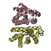
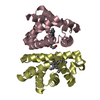
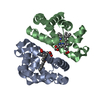
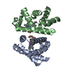
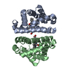
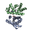
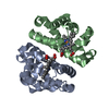
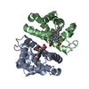
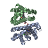
| Title | SCAPHARCA DIMERIC HEMOGLOBIN, MUTANT T72I, DEOXY FORM | ||||||
 Components Components | HEMOGLOBIN | ||||||
 Keywords Keywords | OXYGEN TRANSPORT / HEME / RESPIRATORY PROTEIN | ||||||
| Function / homology |  Function and homology information Function and homology informationoxygen carrier activity / oxygen binding / heme binding / metal ion binding / identical protein binding / cytoplasm Similarity search - Function | ||||||
| Biological species |  Scapharca inaequivalvis (ark clam) Scapharca inaequivalvis (ark clam) | ||||||
| Method |  X-RAY DIFFRACTION / ISOMORPHOUS MOLECULAR REPLACEMENT / Resolution: 1.6 Å X-RAY DIFFRACTION / ISOMORPHOUS MOLECULAR REPLACEMENT / Resolution: 1.6 Å | ||||||
 Authors Authors | Royer Junior, W.E. | ||||||
 Citation Citation | Journal: J.Mol.Biol. / Year: 1998 Title: Mutational destabilization of the critical interface water cluster in Scapharca dimeric hemoglobin: structural basis for altered allosteric activity. Authors: Pardanani, A. / Gambacurta, A. / Ascoli, F. / Royer Jr., W.E. #1: Journal: Proc.Natl.Acad.Sci.USA / Year: 1996Title: Ordered Water Molecules as Key Allosteric Mediators in a Cooperative Dimeric Hemoglobin Authors: Royer Junior, W.E. / Pardanani, A. / Gibson, Q.H. / Peterson, E.S. / Friedman, J.M. #2: Journal: J.Mol.Biol. / Year: 1995Title: A Single Mutation (Thr72-->Ile) at the Subunit Interface is Crucial for the Functional Properties of the Homodimeric Co-Operative Haemoglobin from Scapharca Inaequivalvis Authors: Gambacurta, A. / Piro, M.C. / Coletta, M. / Clementi, M.E. / Polizio, F. / Desideri, A. / Santucci, R. / Ascoli, F. #3: Journal: J.Mol.Biol. / Year: 1994Title: High-Resolution Crystallographic Analysis of a Co-Operative Dimeric Hemoglobin Authors: Royer Junior, W.E. | ||||||
| History |
|
- Structure visualization
| Structure viewer | Molecule: MolmilJmol/JSmol |
|---|
- Downloads & links
-Download
| PDBx/mmCIF format | 4hbi.cif.gz | 74.9 KB | Display | PDBx/mmCIF format |
|---|---|---|---|---|
| PDB format | pdb4hbi.ent.gz | 55.9 KB | Display | PDB format |
| PDBx/mmJSON format | 4hbi.json.gz | Tree view | PDBx/mmJSON format | |
| Others |  Other downloads Other downloads |
-Validation report
| Arichive directory | https://data.pdbj.org/pub/pdb/validation_reports/hb/4hbiftp://data.pdbj.org/pub/pdb/validation_reports/hb/4hbi | HTTPS FTP |
|---|
-Related structure data
| Related structure data |  5hbiC  6hbiC  7hbiC 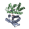 4sdhS S: Starting model for refinement C: citing same article ( |
|---|---|
| Similar structure data |


-Links
 PDBj
PDBj


- Assembly
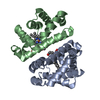
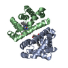
| Deposited unit | 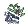
| ||||||||
|---|---|---|---|---|---|---|---|---|---|
| 1 |
| ||||||||
| Unit cell |
| ||||||||
| Noncrystallographic symmetry (NCS) | NCS oper: (Code: given Matrix: (-0.3511, -0.3203, -0.8798), Vector: |
-Components
| #1: Protein | Mass: 15979.356 Da / Num. of mol.: 2 / Mutation: T72I Source method: isolated from a genetically manipulated source Details: SCAPHARCA DIMERIC HEMOGLOBIN, HEME GROUP, PROTOPORPHYRIN IX IRON Source: (gene. exp.) Scapharca inaequivalvis (ark clam) / Plasmid: PGAP1 / Production host:  #2: Chemical |   Mass: 616.487 Da / Num. of mol.: 2 / Source method: obtained synthetically / Formula: C34H32FeN4O4 Mass: 616.487 Da / Num. of mol.: 2 / Source method: obtained synthetically / Formula: C34H32FeN4O4#3: Water | ChemComp-HOH / |  Mass: 18.015 Da / Num. of mol.: 245 / Source method: isolated from a natural source / Formula: H2O Mass: 18.015 Da / Num. of mol.: 245 / Source method: isolated from a natural source / Formula: H2O |
|---|
-Experimental details
-Experiment
| Experiment | Method: X-RAY DIFFRACTION / Number of used crystals: 1 |
|---|
- Sample preparation
| Crystal | Density Matthews: 2.29 Å3/Da / Density % sol: 45 % | |||||||||||||||
|---|---|---|---|---|---|---|---|---|---|---|---|---|---|---|---|---|
| Crystal grow | pH: 8.5 Details: PROTEIN WAS CRYSTALLIZED FROM 2.1M NA/K PHOSPHATE AT PH 8.5 | |||||||||||||||
| Crystal | *PLUS Density % sol: 45 % | |||||||||||||||
| Crystal grow | *PLUS pH: 7.5 / Method: batch method / Details: Royer Junior, W.E., (1994) J.Mol.Biol., 235, 657. | |||||||||||||||
| Components of the solutions | *PLUS
|
-Data collection
| Diffraction | Mean temperature: 293 K |
|---|---|
| Diffraction source | Source: ROTATING ANODE / Type: RIGAKU RUH2R / Wavelength: 1.5418 |
| Detector | Type: RIGAKU / Detector: IMAGE PLATE / Date: Aug 1, 1996 |
| Radiation | Monochromator: GRAPHITE(002) / Monochromatic (M) / Laue (L): M / Scattering type: x-ray |
| Radiation wavelength | Wavelength: 1.5418 Å / Relative weight: 1 |
| Reflection | Resolution: 1.6→20 Å / Num. obs: 35777 / % possible obs: 91.8 % / Observed criterion σ(I): 1 / Redundancy: 4.3 % / Rmerge(I) obs: 0.056 |
| Reflection | *PLUS Num. measured all: 156949 / Rmerge(I) obs: 0.0564 |
- Processing
| Software |
| ||||||||||||||||||||
|---|---|---|---|---|---|---|---|---|---|---|---|---|---|---|---|---|---|---|---|---|---|
| Refinement | Method to determine structure: ISOMORPHOUS MOLECULAR REPLACEMENT Starting model: PDB ENTRY 4SDH Resolution: 1.6→10 Å / Cross valid method: THROUGHOUT / σ(F): 1
| ||||||||||||||||||||
| Refinement step | Cycle: LAST / Resolution: 1.6→10 Å
| ||||||||||||||||||||
| Xplor file |
| ||||||||||||||||||||
| Software | *PLUS Name: X-PLOR / Version: 3.8 / Classification: refinement | ||||||||||||||||||||
| Refine LS restraints | *PLUS
|
