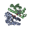
 Movie
Movie Controller


+ Open data

- Basic information
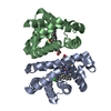
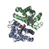
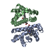
| Entry | Database: PDB / ID: 1nwi | ||||||
|---|---|---|---|---|---|---|---|
| Title | Crystal structure of CO-HbI transformed to an unligated state | ||||||
 Components Components | globin I | ||||||
 Keywords Keywords | OXYGEN STORAGE/TRANSPORT / Invertebrate / hemoglobin / allostery / cooperativity / oxygen-binding / oxygen transport / heme protein / OXYGEN STORAGE-TRANSPORT COMPLEX | ||||||
| Function / homology |  Function and homology information Function and homology informationoxygen carrier activity / oxygen binding / heme binding / metal ion binding / identical protein binding / cytoplasm Similarity search - Function | ||||||
| Biological species |  Scapharca inaequivalvis (ark clam) Scapharca inaequivalvis (ark clam) | ||||||
| Method |  X-RAY DIFFRACTION / MOLECULAR REPLACEMENT / Resolution: 2.5 Å X-RAY DIFFRACTION / MOLECULAR REPLACEMENT / Resolution: 2.5 Å | ||||||
 Authors Authors | Knapp, J.E. / Royer JR., W.E. | ||||||
 Citation Citation | Journal: Biochemistry / Year: 2003 Title: Ligand-linked structural transitions in crystals of a cooperative dimeric hemoglobin. Authors: Knapp, J.E. / Royer Jr., W.E. #1: Journal: J.Mol.Biol. / Year: 1994Title: High resolution crystallographic analysis of a co-operative dimeric hemoglobin Authors: Royer JR., W.E. | ||||||
| History |
|
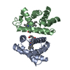
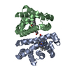
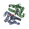
- Structure visualization
| Structure viewer | Molecule: MolmilJmol/JSmol |
|---|
- Downloads & links
-Download
| PDBx/mmCIF format | 1nwi.cif.gz | 117 KB | Display | PDBx/mmCIF format |
|---|---|---|---|---|
| PDB format | pdb1nwi.ent.gz | 94.3 KB | Display | PDB format |
| PDBx/mmJSON format | 1nwi.json.gz | Tree view | PDBx/mmJSON format | |
| Others |  Other downloads Other downloads |
-Validation report
| Arichive directory | https://data.pdbj.org/pub/pdb/validation_reports/nw/1nwiftp://data.pdbj.org/pub/pdb/validation_reports/nw/1nwi | HTTPS FTP |
|---|
-Related structure data
| Related structure data |  1nwnC  1nxfC 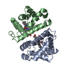 3sdhS S: Starting model for refinement C: citing same article ( |
|---|---|
| Similar structure data |


-Links
 PDBj
PDBj


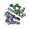
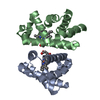
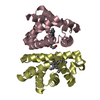
- Assembly
| Deposited unit | 
| ||||||||
|---|---|---|---|---|---|---|---|---|---|
| 1 | 
| ||||||||
| 2 | 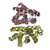
| ||||||||
| Unit cell |
| ||||||||
| Details | The biological assembly is a dimer formed either from the A and B chains or from the C and D chains. |
-Components
| #1: Protein | Mass: 15966.319 Da / Num. of mol.: 4 Source method: isolated from a genetically manipulated source Source: (gene. exp.) Scapharca inaequivalvis (ark clam) / Gene: HbI / Plasmid: PCS-26 / Production host:  #2: Chemical | ChemComp-HEM /   Mass: 616.487 Da / Num. of mol.: 4 / Source method: obtained synthetically / Formula: C34H32FeN4O4 Mass: 616.487 Da / Num. of mol.: 4 / Source method: obtained synthetically / Formula: C34H32FeN4O4#3: Water | ChemComp-HOH / |  Mass: 18.015 Da / Num. of mol.: 148 / Source method: isolated from a natural source / Formula: H2O Mass: 18.015 Da / Num. of mol.: 148 / Source method: isolated from a natural source / Formula: H2O |
|---|
-Experimental details
-Experiment
| Experiment | Method: X-RAY DIFFRACTION / Number of used crystals: 1 |
|---|
- Sample preparation
| Crystal | Density Matthews: 2.18 Å3/Da / Density % sol: 43.06 % |
|---|---|
| Crystal grow | Temperature: 296 K / Method: small tubes / pH: 7.5 Details: Sodium and Potassium phosphate, pH 7.5, SMALL TUBES, temperature 296K |
| Crystal grow | *PLUS Method: unknownDetails: Summerford, C.M., (1995) Protein Eng., 8, 593., Royer, W.E.,Jr. (1994) J. Mol. Biol., 235, 657. |
-Data collection
| Diffraction | Mean temperature: 298 K |
|---|---|
| Diffraction source | Source: ROTATING ANODE / Type: RIGAKU RU200 / Wavelength: 1.5418 Å |
| Detector | Type: RIGAKU RAXIS IIC / Detector: IMAGE PLATE / Date: Jan 29, 2001 / Details: Yale Mirrors |
| Radiation | Monochromator: Yale mirrors / Protocol: SINGLE WAVELENGTH / Monochromatic (M) / Laue (L): M / Scattering type: x-ray |
| Radiation wavelength | Wavelength: 1.5418 Å / Relative weight: 1 |
| Reflection | Resolution: 2.5→40 Å / Num. all: 19646 / Num. obs: 19646 / % possible obs: 95.3 % / Observed criterion σ(F): 1 / Observed criterion σ(I): 1 / Redundancy: 3.8 % / Biso Wilson estimate: 25.7 Å2 / Rmerge(I) obs: 0.077 / Rsym value: 0.077 / Net I/σ(I): 11.5 |
| Reflection shell | Resolution: 2.5→2.59 Å / Rmerge(I) obs: 0.239 / Rsym value: 0.239 / % possible all: 85.6 |
| Reflection | *PLUS Num. measured all: 75147 |
| Reflection shell | *PLUS Highest resolution: 2.5 Å / % possible obs: 85.5 % |
- Processing
| Software |
| |||||||||||||||||||||||||
|---|---|---|---|---|---|---|---|---|---|---|---|---|---|---|---|---|---|---|---|---|---|---|---|---|---|---|
| Refinement | Method to determine structure: MOLECULAR REPLACEMENT Starting model: PDB entry 3SDH Resolution: 2.5→30.95 Å / Rfactor Rfree error: 0.007 / Isotropic thermal model: GROUP / Cross valid method: THROUGHOUT / σ(F): 0 / Stereochemistry target values: Engh & Huber
| |||||||||||||||||||||||||
| Solvent computation | Solvent model: FLAT MODEL / Bsol: 32.9588 Å2 / ksol: 0.320278 e/Å3 | |||||||||||||||||||||||||
| Displacement parameters | Biso mean: 27.9 Å2
| |||||||||||||||||||||||||
| Refine analyze |
| |||||||||||||||||||||||||
| Refinement step | Cycle: LAST / Resolution: 2.5→30.95 Å
| |||||||||||||||||||||||||
| Refine LS restraints |
| |||||||||||||||||||||||||
| LS refinement shell | Resolution: 2.5→2.59 Å / Rfactor Rfree error: 0.026 / Total num. of bins used: 10
| |||||||||||||||||||||||||
| Xplor file |
| |||||||||||||||||||||||||
| Refinement | *PLUS Highest resolution: 2.5 Å / Lowest resolution: 40 Å | |||||||||||||||||||||||||
| Solvent computation | *PLUS | |||||||||||||||||||||||||
| Displacement parameters | *PLUS | |||||||||||||||||||||||||
| Refine LS restraints | *PLUS
|
