 Movie
Movie Controller


+ Open data

- Basic information
| Entry | Database: PDB / ID: 3qib | ||||||
|---|---|---|---|---|---|---|---|
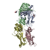
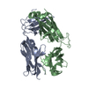
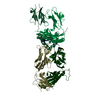
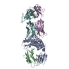
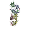
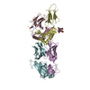
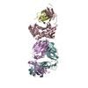
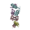
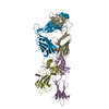
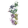
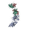
| Title | Crystal structure of the 2B4 TCR in complex with MCC/I-Ek | ||||||
 Components Components |
| ||||||
 Keywords Keywords | IMMUNE SYSTEM / Ig domain | ||||||
| Function / homology |  Function and homology information Function and homology informationPhosphorylation of CD3 and TCR zeta chains / Translocation of ZAP-70 to Immunological synapse / Co-inhibition by PD-1 / MHC class II receptor activity / Generation of second messenger molecules / Downstream TCR signaling / myeloid dendritic cell antigen processing and presentation / antigen processing and presentation of endogenous peptide antigen via MHC class II / regulation of T-helper cell differentiation / positive regulation of CD4-positive, CD25-positive, alpha-beta regulatory T cell differentiation ...Phosphorylation of CD3 and TCR zeta chains / Translocation of ZAP-70 to Immunological synapse / Co-inhibition by PD-1 / MHC class II receptor activity / Generation of second messenger molecules / Downstream TCR signaling / myeloid dendritic cell antigen processing and presentation / antigen processing and presentation of endogenous peptide antigen via MHC class II / regulation of T-helper cell differentiation / positive regulation of CD4-positive, CD25-positive, alpha-beta regulatory T cell differentiation / positive regulation of CD4-positive, alpha-beta T cell activation / antigen processing and presentation of peptide or polysaccharide antigen via MHC class II / MHC class II antigen presentation / positive regulation of memory T cell differentiation / CD4 receptor binding / alpha-beta T cell receptor complex / Translocation of ZAP-70 to Immunological synapse / Phosphorylation of CD3 and TCR zeta chains / immunoglobulin mediated immune response / polysaccharide binding / alpha-beta T cell activation / Generation of second messenger molecules / Co-inhibition by PD-1 / immunological synapse / T cell receptor binding / response to bacterium / respiratory electron transport chain / peptide antigen assembly with MHC class II protein complex / mitochondrial intermembrane space / MHC class II protein complex / positive regulation of T cell mediated cytotoxicity / antigen processing and presentation of exogenous peptide antigen via MHC class II / positive regulation of immune response / peptide antigen binding / cognition / positive regulation of T cell activation / Immunoregulatory interactions between a Lymphoid and a non-Lymphoid cell / MHC class II protein complex binding / T cell receptor signaling pathway / late endosome membrane / Downstream TCR signaling / adaptive immune response / electron transfer activity / lysosome / external side of plasma membrane / lysosomal membrane / heme binding / cell surface / metal ion binding / plasma membrane Similarity search - Function | ||||||
| Biological species |   Homo sapiens (human) Homo sapiens (human) Manduca sexta (tobacco hornworm) Manduca sexta (tobacco hornworm) | ||||||
| Method |  X-RAY DIFFRACTION / SYNCHROTRON / MOLECULAR REPLACEMENT / Resolution: 2.7 Å X-RAY DIFFRACTION / SYNCHROTRON / MOLECULAR REPLACEMENT / Resolution: 2.7 Å | ||||||
 Authors Authors | Ely, L.K. / Newell, E.W. / Davis, M.M. / Garcia, K.C. | ||||||
 Citation Citation | Journal: J.Immunol. / Year: 2011 Title: Structural basis of specificity and cross-reactivity in T cell receptors specific for cytochrome c-I-E(k). Authors: Newell, E.W. / Ely, L.K. / Kruse, A.C. / Reay, P.A. / Rodriguez, S.N. / Lin, A.E. / Kuhns, M.S. / Garcia, K.C. / Davis, M.M. | ||||||
| History |
|
- Structure visualization
| Structure viewer | Molecule: MolmilJmol/JSmol |
|---|
- Downloads & links
-Download
| PDBx/mmCIF format | 3qib.cif.gz | 180.2 KB | Display | PDBx/mmCIF format |
|---|---|---|---|---|
| PDB format | pdb3qib.ent.gz | 139 KB | Display | PDB format |
| PDBx/mmJSON format | 3qib.json.gz | Tree view | PDBx/mmJSON format | |
| Others |  Other downloads Other downloads |
-Validation report
| Arichive directory | https://data.pdbj.org/pub/pdb/validation_reports/qi/3qibftp://data.pdbj.org/pub/pdb/validation_reports/qi/3qib | HTTPS FTP |
|---|
-Related structure data


-Links
 PDBj
PDBj


- Assembly
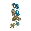
| Deposited unit | 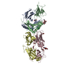
| ||||||||
|---|---|---|---|---|---|---|---|---|---|
| 1 |
| ||||||||
| Unit cell |
|
-Components
-Protein , 4 types, 4 molecules ABCD
| #1: Protein | Mass: 22491.221 Da / Num. of mol.: 1 / Fragment: unp residues 26-216 Source method: isolated from a genetically manipulated source Source: (gene. exp.) Trichoplusia ni (cabbage looper) / References: UniProt: P04224 |
|---|---|
| #2: Protein | Mass: 23344.934 Da / Num. of mol.: 1 / Fragment: unp residues 29-224 Source method: isolated from a genetically manipulated source Source: (gene. exp.) Trichoplusia ni (cabbage looper) / References: UniProt: Q31163, UniProt: P04230*PLUS |
| #3: Protein | Mass: 22807.115 Da / Num. of mol.: 1 Source method: isolated from a genetically manipulated source Source: (gene. exp.) Homo sapiens (human) / Gene: TCRA, TRAC / Production host:  |
| #4: Protein | Mass: 30288.928 Da / Num. of mol.: 1 Source method: isolated from a genetically manipulated source Source: (gene. exp.) |
-Protein/peptide , 1 types, 1 molecules P
| #5: Protein/peptide | Mass: 1393.628 Da / Num. of mol.: 1 / Fragment: unp residues 97-108 Source method: isolated from a genetically manipulated source Source: (gene. exp.) Manduca sexta (tobacco hornworm) / Production host: |
|---|
-Sugars , 3 types, 5 molecules 


| #6: Sugar |  Type: D-saccharide, beta linking / Mass: 221.208 Da / Num. of mol.: 2 Type: D-saccharide, beta linking / Mass: 221.208 Da / Num. of mol.: 2Source method: isolated from a genetically manipulated source Formula: C8H15NO6 #7: Sugar |  Type: L-saccharide, alpha linking / Mass: 164.156 Da / Num. of mol.: 2 Type: L-saccharide, alpha linking / Mass: 164.156 Da / Num. of mol.: 2Source method: isolated from a genetically manipulated source Formula: C6H12O5 #9: Sugar | ChemComp-BMA / |  Type: D-saccharide, beta linking / Mass: 180.156 Da / Num. of mol.: 1 Type: D-saccharide, beta linking / Mass: 180.156 Da / Num. of mol.: 1Source method: isolated from a genetically manipulated source Formula: C6H12O6 |
|---|
-Non-polymers , 2 types, 113 molecules 

| #8: Chemical | ChemComp-PEG /  Mass: 106.120 Da / Num. of mol.: 7 / Source method: obtained synthetically / Formula: C4H10O3 Mass: 106.120 Da / Num. of mol.: 7 / Source method: obtained synthetically / Formula: C4H10O3#10: Water | ChemComp-HOH / | Mass: 18.015 Da / Num. of mol.: 106 / Source method: isolated from a natural source / Formula: H2O |
|---|
-Details
| Has protein modification | Y |
|---|
-Experimental details
-Experiment
| Experiment | Method: X-RAY DIFFRACTION / Number of used crystals: 1 |
|---|
- Sample preparation
| Crystal | Density Matthews: 3.97 Å3/Da / Density % sol: 68.99 % |
|---|---|
| Crystal grow | Temperature: 295 K / pH: 6.2 Details: 50% PEG200, 0.2 M sodium chloride, 0.1 M sodium potassium phosphate, pH 6.2, VAPOR DIFFUSION, temperature 295K |
-Data collection
| Diffraction | Mean temperature: 100 K |
|---|---|
| Diffraction source | Source: SYNCHROTRON / Site: ALS  / Beamline: 8.2.1 / Wavelength: 1 / Beamline: 8.2.1 / Wavelength: 1 |
| Detector | Type: ADSC QUANTUM 315r / Detector: CCD / Date: Oct 11, 2009 |
| Radiation | Monochromator: SI 111 CHANNEL / Protocol: SINGLE WAVELENGTH / Monochromatic (M) / Laue (L): M / Scattering type: x-ray |
| Radiation wavelength | Wavelength: 1 Å / Relative weight: 1 |
| Reflection | Resolution: 2.4→50 Å / Num. obs: 43820 |
- Processing
| Software | Name: PHENIX / Version: (phenix.refine: 1.6.2_432) / Classification: refinement | |||||||||||||||||||||||||||||||||||||||||||||||||||||||||||||||||||||||||||||
|---|---|---|---|---|---|---|---|---|---|---|---|---|---|---|---|---|---|---|---|---|---|---|---|---|---|---|---|---|---|---|---|---|---|---|---|---|---|---|---|---|---|---|---|---|---|---|---|---|---|---|---|---|---|---|---|---|---|---|---|---|---|---|---|---|---|---|---|---|---|---|---|---|---|---|---|---|---|---|
| Refinement | Method to determine structure: MOLECULAR REPLACEMENT / Resolution: 2.7→34.97 Å / SU ML: 0.34 / σ(F): 1.34 / Phase error: 24.87 / Stereochemistry target values: ML
| |||||||||||||||||||||||||||||||||||||||||||||||||||||||||||||||||||||||||||||
| Solvent computation | Shrinkage radii: 0.95 Å / VDW probe radii: 1.2 Å / Solvent model: FLAT BULK SOLVENT MODEL / Bsol: 34.63 Å2 / ksol: 0.34 e/Å3 | |||||||||||||||||||||||||||||||||||||||||||||||||||||||||||||||||||||||||||||
| Displacement parameters |
| |||||||||||||||||||||||||||||||||||||||||||||||||||||||||||||||||||||||||||||
| Refinement step | Cycle: LAST / Resolution: 2.7→34.97 Å
| |||||||||||||||||||||||||||||||||||||||||||||||||||||||||||||||||||||||||||||
| Refine LS restraints |
| |||||||||||||||||||||||||||||||||||||||||||||||||||||||||||||||||||||||||||||
| LS refinement shell |
|
