Protocol: SINGLE WAVELENGTH / Monochromatic (M) / Laue (L): M / Scattering type: x-ray
Radiation wavelength
Wavelength: 1.5 Å / Relative weight: 1
Reflection
Redundancy: 5.6 % / Av σ(I) over netI: 7.4 / Number: 124052 / Rsym value: 0.101 / D res high: 2.2 Å / D res low: 37.484 Å / Num. obs: 22340 / % possible obs: 100
Diffraction reflection shell
Highest resolution (Å)
Lowest resolution (Å)
% possible obs (%)
ID
Rmerge(I) obs
Rsym value
Redundancy
6.96
37.48
99.4
1
0.025
0.025
5.4
4.92
6.96
100
1
0.038
0.038
5.6
4.02
4.92
100
1
0.037
0.037
5.6
3.48
4.02
100
1
0.054
0.054
5.6
3.11
3.48
100
1
0.1
0.1
5.6
2.84
3.11
100
1
0.179
0.179
5.6
2.63
2.84
100
1
0.291
0.291
5.6
2.46
2.63
100
1
0.431
0.431
5.5
2.32
2.46
100
1
0.61
0.61
5.5
2.2
2.32
100
1
0.851
0.851
5.5
Reflection
Resolution: 2.2→37.484 Å / Num. all: 22340 / Num. obs: 22340 / % possible obs: 100 % / Redundancy: 5.6 % / Biso Wilson estimate: 39.9 Å2 / Rmerge(I) obs: 0.101 / Rsym value: 0.101 / Net I/σ(I): 13.3
Reflection shell
Resolution (Å)
Redundancy (%)
Rmerge(I) obs
Mean I/σ(I) obs
Num. measured all
Num. unique all
Rsym value
% possible all
2.2-2.32
5.5
0.851
0.9
17791
3251
0.851
100
2.32-2.46
5.5
0.61
1.3
16914
3076
0.61
100
2.46-2.63
5.5
0.431
1.8
15875
2869
0.431
100
2.63-2.84
5.6
0.291
2.7
14902
2684
0.291
100
2.84-3.11
5.6
0.179
4.3
13930
2492
0.179
100
3.11-3.48
5.6
0.1
7.6
12619
2247
0.1
100
3.48-4.02
5.6
0.054
14
11156
1978
0.054
100
4.02-4.92
5.6
0.037
19.3
9544
1695
0.037
100
4.92-6.96
5.6
0.038
19.2
7353
1309
0.038
100
6.96-37.484
5.4
0.025
24.7
3968
739
0.025
99.4
-
Phasing
Phasing
Method: molecular replacement
Phasing MR
Rfactor: 32.72 / Model details: Phaser MODE: MR_AUTO
Resolution: 2.2→37.48 Å / Cor.coef. Fo:Fc: 0.954 / Cor.coef. Fo:Fc free: 0.933 / WRfactor Rfree: 0.1957 / WRfactor Rwork: 0.1639 / Occupancy max: 1 / Occupancy min: 0.5 / FOM work R set: 0.8819 / SU B: 9.776 / SU ML: 0.118 / SU R Cruickshank DPI: 0.1943 / SU Rfree: 0.1733 / Cross valid method: THROUGHOUT / σ(F): 0 / ESU R Free: 0.173 / Stereochemistry target values: MAXIMUM LIKELIHOOD Details: HYDROGENS HAVE BEEN ADDED IN THE RIDING POSITIONS U VALUES: WITH TLS ADDED
Rfactor
Num. reflection
% reflection
Selection details
Rfree
0.2268
1110
5 %
RANDOM
Rwork
0.1858
-
-
-
all
0.1878
22314
-
-
obs
0.1878
22310
99.98 %
-
Solvent computation
Ion probe radii: 0.8 Å / Shrinkage radii: 0.8 Å / VDW probe radii: 1.4 Å / Solvent model: MASK
In the structure databanks used in Yorodumi, some data are registered as the other names, "COVID-19 virus" and "2019-nCoV". Here are the details of the virus and the list of structure data.
Jan 31, 2019. EMDB accession codes are about to change! (news from PDBe EMDB page)
EMDB accession codes are about to change! (news from PDBe EMDB page)
The allocation of 4 digits for EMDB accession codes will soon come to an end. Whilst these codes will remain in use, new EMDB accession codes will include an additional digit and will expand incrementally as the available range of codes is exhausted. The current 4-digit format prefixed with “EMD-” (i.e. EMD-XXXX) will advance to a 5-digit format (i.e. EMD-XXXXX), and so on. It is currently estimated that the 4-digit codes will be depleted around Spring 2019, at which point the 5-digit format will come into force.
The EM Navigator/Yorodumi systems omit the EMD- prefix.
Related info.:Q: What is EMD? / ID/Accession-code notation in Yorodumi/EM Navigator
Yorodumi is a browser for structure data from EMDB, PDB, SASBDB, etc.
This page is also the successor to EM Navigator detail page, and also detail information page/front-end page for Omokage search.
The word "yorodu" (or yorozu) is an old Japanese word meaning "ten thousand". "mi" (miru) is to see.
Related info.:EMDB / PDB / SASBDB / Comparison of 3 databanks / Yorodumi Search / Aug 31, 2016. New EM Navigator & Yorodumi / Yorodumi Papers / Jmol/JSmol / Function and homology information / Changes in new EM Navigator and Yorodumi
 Movie
Movie Controller

 Yorodumi
 Open data
 Basic information
 Components
 Keywords
 Function and homology information
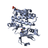
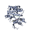
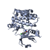
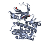
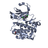
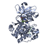
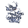
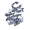
 Homo sapiens (human)
 X-RAY DIFFRACTION /
  Authors
 Citation
 Structure visualization
 Downloads & links
 Other downloads
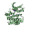


 PDBj
PDBj


 Assembly
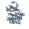

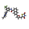


 Mass: 634.668 Da / Num. of mol.: 1 / Source method: obtained synthetically / Formula: C33H29F3N4O4S
 Mass: 22.990 Da / Num. of mol.: 1 / Source method: obtained synthetically / Formula: Na
 Mass: 35.453 Da / Num. of mol.: 1 / Source method: obtained synthetically / Formula: Cl
 Sample preparation
 Processing