 Movie
Movie Controller

[English] 日本語
 Yorodumi
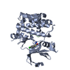
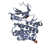
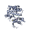
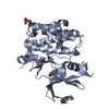
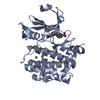
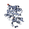
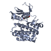
- PDB-4gw8: Human proto-oncogene serine threonine kinase (PIM1) in complex wi... -
+ Open data

- Basic information
| Entry | Database: PDB / ID: 4gw8 | ||||||
|---|---|---|---|---|---|---|---|
| Title | Human proto-oncogene serine threonine kinase (PIM1) in complex with a consensus peptide and Leucettine L41 | ||||||
 Components Components |
| ||||||
 Keywords Keywords | TRANSFERASE / ONCOGENE / KINASE / SERINE-THREONINE / PIM1 / STRUCTURAL GENOMICS CONSORTIUM / SGC / ALTERNATIVE INITIATION / ATP-BINDING / MANGANESE / MEMBRANE / METAL-BINDING / NUCLEOTIDE-BINDING / NUCLEUS / PHOSPHOPROTEIN / PROTO-ONCOGENE / SERINE/THREONINE-PROTEIN KINASE / HOST-VIRUS INTERACTION / VIRAL IMMUNOEVASION / VIRION / VIRULENCE / CELL CYCLE / CELL MEMBRANE | ||||||
| Function / homology |  Function and homology information Function and homology informationregulation of transmembrane transporter activity / positive regulation of cardioblast proliferation / positive regulation of cyclin-dependent protein serine/threonine kinase activity / regulation of hematopoietic stem cell proliferation / vitamin D receptor signaling pathway / cellular detoxification / STAT5 activation downstream of FLT3 ITD mutants / transcription factor binding / positive regulation of protein serine/threonine kinase activity / ribosomal small subunit binding ...regulation of transmembrane transporter activity / positive regulation of cardioblast proliferation / positive regulation of cyclin-dependent protein serine/threonine kinase activity / regulation of hematopoietic stem cell proliferation / vitamin D receptor signaling pathway / cellular detoxification / STAT5 activation downstream of FLT3 ITD mutants / transcription factor binding / positive regulation of protein serine/threonine kinase activity / ribosomal small subunit binding / : / positive regulation of cardiac muscle cell proliferation / positive regulation of brown fat cell differentiation / positive regulation of TORC1 signaling / Signaling by FLT3 fusion proteins / regulation of mitotic cell cycle / negative regulation of innate immune response / protein serine/threonine kinase activator activity / cellular response to type II interferon / protein autophosphorylation / manganese ion binding / Interleukin-4 and Interleukin-13 signaling / protein phosphorylation / non-specific serine/threonine protein kinase / protein stabilization / protein serine kinase activity / protein serine/threonine kinase activity / apoptotic process / negative regulation of apoptotic process / nucleolus / positive regulation of DNA-templated transcription / nucleoplasm / ATP binding / nucleus / plasma membrane / cytosol / cytoplasm Similarity search - Function | ||||||
| Biological species |  Homo sapiens (human) Homo sapiens (human) | ||||||
| Method |  X-RAY DIFFRACTION / MOLECULAR REPLACEMENT / molecular replacement / Resolution: 2 Å X-RAY DIFFRACTION / MOLECULAR REPLACEMENT / molecular replacement / Resolution: 2 Å | ||||||
 Authors Authors | Filippakopoulos, P. / Bullock, A. / von Delft, F. / Bountra, C. / Arrowsmith, C.H. / Edwards, A. / Meijer, L. / Knapp, S. / Structural Genomics Consortium (SGC) | ||||||
 Citation Citation | Journal: J.Med.Chem. / Year: 2012 Title: Selectivity, cocrystal structures, and neuroprotective properties of leucettines, a family of protein kinase inhibitors derived from the marine sponge alkaloid leucettamine B. Authors: Tahtouh, T. / Elkins, J.M. / Filippakopoulos, P. / Soundararajan, M. / Burgy, G. / Durieu, E. / Cochet, C. / Schmid, R.S. / Lo, D.C. / Delhommel, F. / Oberholzer, A.E. / Pearl, L.H. / ...Authors: Tahtouh, T. / Elkins, J.M. / Filippakopoulos, P. / Soundararajan, M. / Burgy, G. / Durieu, E. / Cochet, C. / Schmid, R.S. / Lo, D.C. / Delhommel, F. / Oberholzer, A.E. / Pearl, L.H. / Carreaux, F. / Bazureau, J.P. / Knapp, S. / Meijer, L. | ||||||
| History |
|
- Structure visualization
| Structure viewer | Molecule: MolmilJmol/JSmol |
|---|
- Downloads & links
-Download
| PDBx/mmCIF format | 4gw8.cif.gz | 136.3 KB | Display | PDBx/mmCIF format |
|---|---|---|---|---|
| PDB format | pdb4gw8.ent.gz | 104.6 KB | Display | PDB format |
| PDBx/mmJSON format | 4gw8.json.gz | Tree view | PDBx/mmJSON format | |
| Others |  Other downloads Other downloads |
-Validation report
| Arichive directory | https://data.pdbj.org/pub/pdb/validation_reports/gw/4gw8ftp://data.pdbj.org/pub/pdb/validation_reports/gw/4gw8 | HTTPS FTP |
|---|
-Related structure data
| Related structure data |  4azeC  4azfC  4b7tC  2c3iS S: Starting model for refinement C: citing same article ( |
|---|---|
| Similar structure data |


-Links
 PDBj
PDBj

- Assembly
| Deposited unit | 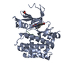
| ||||||||
|---|---|---|---|---|---|---|---|---|---|
| 1 |
| ||||||||
| Unit cell |
|
-Components
| #1: Protein | Mass: 35590.500 Da / Num. of mol.: 1 / Fragment: Isoform 2 / Mutation: R250G Source method: isolated from a genetically manipulated source Details: THE EXPRESSED PROTEIN HAS THE SEQUENCE FROM GI33304198 WHICH DIFFERS FROM GI 4505811 BY A SINGLE MUTATION R250G Source: (gene. exp.) Homo sapiens (human) / Gene: PIM1 / Plasmid: pNIC28-Bsa4 / Production host:  References: UniProt: P11309, non-specific serine/threonine protein kinase |
|---|---|
| #2: Protein/peptide | Mass: 1592.850 Da / Num. of mol.: 1 / Source method: obtained synthetically / Details: Synthetic consensus peptide (Pimtide) |
| #3: Chemical | ChemComp-3RA / 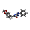  Mass: 307.303 Da / Num. of mol.: 1 / Source method: obtained synthetically / Formula: C17H13N3O3 Mass: 307.303 Da / Num. of mol.: 1 / Source method: obtained synthetically / Formula: C17H13N3O3 |
| #4: Chemical | ChemComp-EDO /   Mass: 62.068 Da / Num. of mol.: 1 / Source method: obtained synthetically / Formula: C2H6O2 Mass: 62.068 Da / Num. of mol.: 1 / Source method: obtained synthetically / Formula: C2H6O2 |
| #5: Water | ChemComp-HOH /  Mass: 18.015 Da / Num. of mol.: 198 / Source method: isolated from a natural source / Formula: H2O Mass: 18.015 Da / Num. of mol.: 198 / Source method: isolated from a natural source / Formula: H2O |
-Experimental details
-Experiment
| Experiment | Method: X-RAY DIFFRACTION / Number of used crystals: 1 |
|---|
- Sample preparation
| Crystal | Density Matthews: 3 Å3/Da / Density % sol: 59.02 % |
|---|---|
| Crystal grow | Temperature: 277 K / Method: vapor diffusion, sitting drop / pH: 8.5 Details: 0.16M Na(OAc), 0.08M BTProp pH 8.5 , 16% PEG3350, 8% EtGly, VAPOR DIFFUSION, SITTING DROP, temperature 277K |
-Data collection
| Diffraction | Mean temperature: 100 K | ||||||||||||||||||||||||||||||||||||||||||||||||||||||||||||||||||||||||||||||||||||||||
|---|---|---|---|---|---|---|---|---|---|---|---|---|---|---|---|---|---|---|---|---|---|---|---|---|---|---|---|---|---|---|---|---|---|---|---|---|---|---|---|---|---|---|---|---|---|---|---|---|---|---|---|---|---|---|---|---|---|---|---|---|---|---|---|---|---|---|---|---|---|---|---|---|---|---|---|---|---|---|---|---|---|---|---|---|---|---|---|---|---|
| Diffraction source | Source: ROTATING ANODE / Type: RIGAKU FR-E SUPERBRIGHT / Wavelength: 1.5418 Å | ||||||||||||||||||||||||||||||||||||||||||||||||||||||||||||||||||||||||||||||||||||||||
| Detector | Type: RIGAKU RAXIS IV / Detector: IMAGE PLATE / Date: Oct 28, 2008 | ||||||||||||||||||||||||||||||||||||||||||||||||||||||||||||||||||||||||||||||||||||||||
| Radiation | Protocol: SINGLE WAVELENGTH / Monochromatic (M) / Laue (L): M / Scattering type: x-ray | ||||||||||||||||||||||||||||||||||||||||||||||||||||||||||||||||||||||||||||||||||||||||
| Radiation wavelength | Wavelength: 1.5418 Å / Relative weight: 1 | ||||||||||||||||||||||||||||||||||||||||||||||||||||||||||||||||||||||||||||||||||||||||
| Reflection | Redundancy: 6.3 % / Av σ(I) over netI: 9.7 / Number: 180841 / Rsym value: 0.074 / D res high: 1.9 Å / D res low: 19.582 Å / Num. obs: 28862 / % possible obs: 97 | ||||||||||||||||||||||||||||||||||||||||||||||||||||||||||||||||||||||||||||||||||||||||
| Diffraction reflection shell |
| ||||||||||||||||||||||||||||||||||||||||||||||||||||||||||||||||||||||||||||||||||||||||
| Reflection | Resolution: 2→19.582 Å / Num. all: 29755 / Num. obs: 28862 / % possible obs: 97 % / Redundancy: 6.3 % / Biso Wilson estimate: 34.5 Å2 / Rmerge(I) obs: 0.074 / Rsym value: 0.074 / Net I/σ(I): 17.7 | ||||||||||||||||||||||||||||||||||||||||||||||||||||||||||||||||||||||||||||||||||||||||
| Reflection shell | Diffraction-ID: 1
|
-Phasing
| Phasing | Method: molecular replacement | |||||||||
|---|---|---|---|---|---|---|---|---|---|---|
| Phasing MR | Rfactor: 31.57 / Model details: Phaser MODE: MR_AUTO
|
- Processing
| Software |
| ||||||||||||||||||||||||||||||||||||||||||||||||||||||||||||||||||||||||||||||||||||||||||||||||||||
|---|---|---|---|---|---|---|---|---|---|---|---|---|---|---|---|---|---|---|---|---|---|---|---|---|---|---|---|---|---|---|---|---|---|---|---|---|---|---|---|---|---|---|---|---|---|---|---|---|---|---|---|---|---|---|---|---|---|---|---|---|---|---|---|---|---|---|---|---|---|---|---|---|---|---|---|---|---|---|---|---|---|---|---|---|---|---|---|---|---|---|---|---|---|---|---|---|---|---|---|---|---|
| Refinement | Method to determine structure: MOLECULAR REPLACEMENT Starting model: PDB ENTRY 2C3I Resolution: 2→19.58 Å / Cor.coef. Fo:Fc: 0.962 / Cor.coef. Fo:Fc free: 0.943 / WRfactor Rfree: 0.2218 / WRfactor Rwork: 0.1977 / Occupancy max: 1 / Occupancy min: 0.3 / FOM work R set: 0.8844 / SU B: 6.932 / SU ML: 0.089 / SU R Cruickshank DPI: 0.1725 / SU Rfree: 0.152 / Cross valid method: THROUGHOUT / σ(F): 0 / ESU R: 0.139 / ESU R Free: 0.133 / Stereochemistry target values: MAXIMUM LIKELIHOOD Details: HYDROGENS HAVE BEEN ADDED IN THE RIDING POSITIONS U VALUES: RESIDUAL ONLY
| ||||||||||||||||||||||||||||||||||||||||||||||||||||||||||||||||||||||||||||||||||||||||||||||||||||
| Solvent computation | Ion probe radii: 0.8 Å / Shrinkage radii: 0.8 Å / VDW probe radii: 1.2 Å / Solvent model: MASK | ||||||||||||||||||||||||||||||||||||||||||||||||||||||||||||||||||||||||||||||||||||||||||||||||||||
| Displacement parameters | Biso max: 78.14 Å2 / Biso mean: 36.226 Å2 / Biso min: 2 Å2
| ||||||||||||||||||||||||||||||||||||||||||||||||||||||||||||||||||||||||||||||||||||||||||||||||||||
| Refinement step | Cycle: LAST / Resolution: 2→19.58 Å
| ||||||||||||||||||||||||||||||||||||||||||||||||||||||||||||||||||||||||||||||||||||||||||||||||||||
| Refine LS restraints |
| ||||||||||||||||||||||||||||||||||||||||||||||||||||||||||||||||||||||||||||||||||||||||||||||||||||
| LS refinement shell | Resolution: 2→2.051 Å / Total num. of bins used: 20
| ||||||||||||||||||||||||||||||||||||||||||||||||||||||||||||||||||||||||||||||||||||||||||||||||||||
| Refinement TLS params. | Method: refined / Refine-ID: X-RAY DIFFRACTION
| ||||||||||||||||||||||||||||||||||||||||||||||||||||||||||||||||||||||||||||||||||||||||||||||||||||
| Refinement TLS group |
|
