Mass: 18.015 Da / Num. of mol.: 161 / Source method: isolated from a natural source / Formula: H2O
Sequence details
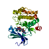
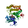
AUTHORS STATE THAT THE EXPRESSED PROTEIN HAS THE SEQUENCE FROM GI 33304198 WHICH DIFFERS FROM GI ...AUTHORS STATE THAT THE EXPRESSED PROTEIN HAS THE SEQUENCE FROM GI 33304198 WHICH DIFFERS FROM GI 4505811 BY A SINGLE MUTATION R250G. AUTHORS CAN NO LONGER FIND GI|33304198 ON THE DATABASES. AUTHORS ARE NOT SURE IF THIS IS DUE TO POLYMORPHISM OR NOT. AUTHORS CONFIRM BY ESI THAT THE SEQUENCE WE HAVE CORRESPONDS TO THE R250G MUTANT AND ALSO IN OUR EXPERIMENTAL DENSITY WE SEE A G AND NOT A LARGERAMINOACID.
-
Experimental details
-
Experiment
Experiment
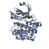
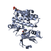
Method: X-RAY DIFFRACTION / Number of used crystals: 1
-
Sample preparation
Crystal
Density Matthews: 3 Å3/Da / Density % sol: 58.99 %
Crystal grow
Temperature: 277 K / Method: vapor diffusion, sitting drop / pH: 8.5 Details: 0.2M NaNO3 0.1M BTProp pH 8.5 20% PEG3350 10% EtGly, VAPOR DIFFUSION, SITTING DROP, temperature 277K
In the structure databanks used in Yorodumi, some data are registered as the other names, "COVID-19 virus" and "2019-nCoV". Here are the details of the virus and the list of structure data.
Jan 31, 2019. EMDB accession codes are about to change! (news from PDBe EMDB page)
EMDB accession codes are about to change! (news from PDBe EMDB page)
The allocation of 4 digits for EMDB accession codes will soon come to an end. Whilst these codes will remain in use, new EMDB accession codes will include an additional digit and will expand incrementally as the available range of codes is exhausted. The current 4-digit format prefixed with “EMD-” (i.e. EMD-XXXX) will advance to a 5-digit format (i.e. EMD-XXXXX), and so on. It is currently estimated that the 4-digit codes will be depleted around Spring 2019, at which point the 5-digit format will come into force.
The EM Navigator/Yorodumi systems omit the EMD- prefix.
Related info.:Q: What is EMD? / ID/Accession-code notation in Yorodumi/EM Navigator
Yorodumi is a browser for structure data from EMDB, PDB, SASBDB, etc.
This page is also the successor to EM Navigator detail page, and also detail information page/front-end page for Omokage search.
The word "yorodu" (or yorozu) is an old Japanese word meaning "ten thousand". "mi" (miru) is to see.
Related info.:EMDB / PDB / SASBDB / Comparison of 3 databanks / Yorodumi Search / Aug 31, 2016. New EM Navigator & Yorodumi / Yorodumi Papers / Jmol/JSmol / Function and homology information / Changes in new EM Navigator and Yorodumi
 Movie
Movie Controller

 Yorodumi
 Open data
 Basic information
 Components
 Keywords
 Function and homology information
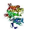
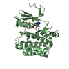
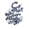
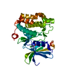
 Homo sapiens (human)
 X-RAY DIFFRACTION /
  Authors
 Citation
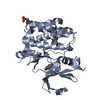
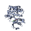
 Structure visualization
 Downloads & links
 Other downloads
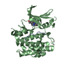


 PDBj
PDBj

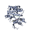
 Assembly

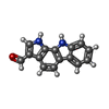
 Mass: 234.253 Da / Num. of mol.: 1 / Source method: obtained synthetically / Formula: C15H10N2O
 Mass: 18.015 Da / Num. of mol.: 161 / Source method: isolated from a natural source / Formula: H2O
 Sample preparation
 Processing