 Movie
Movie Controller

[English] 日本語
 Yorodumi
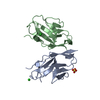
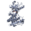
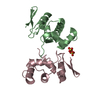
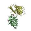
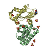
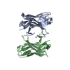
- PDB-3qe1: Crystal Structure of PDZ domain of sorting nexin 27 (SNX27) fused... -
+ Open data

- Basic information
| Entry | Database: PDB / ID: 3qe1 | ||||||
|---|---|---|---|---|---|---|---|
| Title | Crystal Structure of PDZ domain of sorting nexin 27 (SNX27) fused to the C-terminal residues (ESESKV) of GIRK3 | ||||||
 Components Components | Sorting nexin-27, G protein-activated inward rectifier potassium channel 3 chimera | ||||||
 Keywords Keywords | PROTEIN BINDING / PDZ domain / PDZ binding / GIRK3 regulation / early endosomes / brain / neurons | ||||||
| Function / homology |  Function and homology information Function and homology informationneurotransmitter receptor transport to plasma membrane / postsynaptic early endosome / G-protein activated inward rectifier potassium channel activity / establishment of protein localization to plasma membrane / regulation of presynaptic membrane potential / positive regulation of AMPA glutamate receptor clustering / postsynaptic recycling endosome / inward rectifier potassium channel complex / WASH complex / retromer complex ...neurotransmitter receptor transport to plasma membrane / postsynaptic early endosome / G-protein activated inward rectifier potassium channel activity / establishment of protein localization to plasma membrane / regulation of presynaptic membrane potential / positive regulation of AMPA glutamate receptor clustering / postsynaptic recycling endosome / inward rectifier potassium channel complex / WASH complex / retromer complex / response to methamphetamine hydrochloride / endosome to plasma membrane protein transport / phosphatidylinositol-3-phosphate binding / endocytic recycling / regulation of synapse maturation / Activation of G protein gated Potassium channels / Inhibition of voltage gated Ca2+ channels via Gbeta/gamma subunits / endosomal transport / endosome to lysosome transport / potassium ion import across plasma membrane / parallel fiber to Purkinje cell synapse / immunological synapse / regulation of postsynaptic membrane neurotransmitter receptor levels / phosphatidylinositol binding / ionotropic glutamate receptor binding / PDZ domain binding / cellular response to nerve growth factor stimulus / intracellular protein transport / Schaffer collateral - CA1 synapse / calcium-dependent protein binding / nervous system development / presynaptic membrane / early endosome membrane / early endosome / postsynapse / endosome / glutamatergic synapse / signal transduction / plasma membrane / cytosol Similarity search - Function | ||||||
| Biological species |  | ||||||
| Method |  X-RAY DIFFRACTION / MOLECULAR REPLACEMENT / Resolution: 1.68 Å X-RAY DIFFRACTION / MOLECULAR REPLACEMENT / Resolution: 1.68 Å | ||||||
 Authors Authors | Balana, B. / Kwiatkowski, W. / Choe, S. | ||||||
 Citation Citation | Journal: Proc.Natl.Acad.Sci.USA / Year: 2011 Title: Mechanism underlying selective regulation of G protein-gated inwardly rectifying potassium channels by the psychostimulant-sensitive sorting nexin 27. Authors: Balana, B. / Maslennikov, I. / Kwiatkowski, W. / Stern, K.M. / Bahima, L. / Choe, S. / Slesinger, P.A. | ||||||
| History |
|
- Structure visualization
| Structure viewer | Molecule: MolmilJmol/JSmol |
|---|
- Downloads & links
-Download
| PDBx/mmCIF format | 3qe1.cif.gz | 34.9 KB | Display | PDBx/mmCIF format |
|---|---|---|---|---|
| PDB format | pdb3qe1.ent.gz | 23.5 KB | Display | PDB format |
| PDBx/mmJSON format | 3qe1.json.gz | Tree view | PDBx/mmJSON format | |
| Others |  Other downloads Other downloads |
-Validation report
| Arichive directory | https://data.pdbj.org/pub/pdb/validation_reports/qe/3qe1ftp://data.pdbj.org/pub/pdb/validation_reports/qe/3qe1 | HTTPS FTP |
|---|
-Related structure data
| Related structure data | 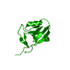 3qdoC 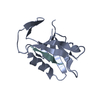 3qglC  2ocsS C: citing same article ( S: Starting model for refinement |
|---|---|
| Similar structure data |


-Links
 PDBj
PDBj
- Assembly
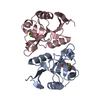
| Deposited unit | 
| ||||||||
|---|---|---|---|---|---|---|---|---|---|
| 1 | 
| ||||||||
| Unit cell |
|
-Components
| #1: Protein | Mass: 11230.646 Da / Num. of mol.: 1 Fragment: PDZ domain (UNP residues 39-133), GIRK-3 C-terminus (UNP residues 388-393) Source method: isolated from a genetically manipulated source Source: (gene. exp.)  |
|---|---|
| #2: Water | ChemComp-HOH /  Mass: 18.015 Da / Num. of mol.: 120 / Source method: isolated from a natural source / Formula: H2O Mass: 18.015 Da / Num. of mol.: 120 / Source method: isolated from a natural source / Formula: H2O |
| Sequence details | CHAIN A IS A CHIMERA: SNX27 PDZ DOMAIN (UNP RESIDUES 39-133), GIRK-3 C-TERMINUS (UNP RESIDUES 388-393) |
-Experimental details
-Experiment
| Experiment | Method: X-RAY DIFFRACTION / Number of used crystals: 1 |
|---|
- Sample preparation
| Crystal | Density Matthews: 2.09 Å3/Da / Density % sol: 41.11 % |
|---|---|
| Crystal grow | Temperature: 298 K / Method: vapor diffusion, hanging drop / pH: 7.5 Details: 0.2 M calcium chloride, 0.1 M HEPES, 28% v/v PEG400, pH 7.5, VAPOR DIFFUSION, HANGING DROP, temperature 298K |
-Data collection
| Diffraction | Mean temperature: 100 K |
|---|---|
| Diffraction source | Source: ROTATING ANODE / Type: RIGAKU MICROMAX-007 / Wavelength: 1.5418 |
| Detector | Type: RIGAKU RAXIS IV++ / Detector: IMAGE PLATE / Date: Aug 26, 2008 / Details: mirrors |
| Radiation | Monochromator: mirrors / Protocol: SINGLE WAVELENGTH / Monochromatic (M) / Laue (L): M / Scattering type: x-ray |
| Radiation wavelength | Wavelength: 1.5418 Å / Relative weight: 1 |
| Reflection | Resolution: 1.68→50 Å / Num. obs: 8921 / % possible obs: 86.3 % / Observed criterion σ(F): 0 / Redundancy: 4.8 % / Biso Wilson estimate: 39.25 Å2 / Rmerge(I) obs: 0.039 / Net I/σ(I): 39.53 |
| Reflection shell | Resolution: 1.68→1.71 Å / Redundancy: 3.3 % / Rmerge(I) obs: 0.174 / Mean I/σ(I) obs: 6.68 / Num. unique all: 232 / % possible all: 45.2 |
- Processing
| Software |
| ||||||||||||||||||||||||||||||||||||||||||||||||||||||||||||||||||||||||||||||||||||||||||||||||||||||||||||||||||||||||||||||||||||||||||||||||||||||||||||||||||||||||||
|---|---|---|---|---|---|---|---|---|---|---|---|---|---|---|---|---|---|---|---|---|---|---|---|---|---|---|---|---|---|---|---|---|---|---|---|---|---|---|---|---|---|---|---|---|---|---|---|---|---|---|---|---|---|---|---|---|---|---|---|---|---|---|---|---|---|---|---|---|---|---|---|---|---|---|---|---|---|---|---|---|---|---|---|---|---|---|---|---|---|---|---|---|---|---|---|---|---|---|---|---|---|---|---|---|---|---|---|---|---|---|---|---|---|---|---|---|---|---|---|---|---|---|---|---|---|---|---|---|---|---|---|---|---|---|---|---|---|---|---|---|---|---|---|---|---|---|---|---|---|---|---|---|---|---|---|---|---|---|---|---|---|---|---|---|---|---|---|---|---|---|---|
| Refinement | Method to determine structure: MOLECULAR REPLACEMENT Starting model: PDB ENTRY 2OCS Resolution: 1.68→37.9 Å / Cor.coef. Fo:Fc: 0.97 / Cor.coef. Fo:Fc free: 0.932 / SU B: 2.178 / SU ML: 0.074 / Cross valid method: THROUGHOUT / σ(F): 0 / ESU R: 0.118 / ESU R Free: 0.127 / Stereochemistry target values: MAXIMUM LIKELIHOOD Details: HYDROGENS HAVE BEEN ADDED IN THE RIDING POSITIONS U VALUES: REFINED INDIVIDUALLY
| ||||||||||||||||||||||||||||||||||||||||||||||||||||||||||||||||||||||||||||||||||||||||||||||||||||||||||||||||||||||||||||||||||||||||||||||||||||||||||||||||||||||||||
| Solvent computation | Ion probe radii: 0.8 Å / Shrinkage radii: 0.8 Å / VDW probe radii: 1.4 Å / Solvent model: MASK | ||||||||||||||||||||||||||||||||||||||||||||||||||||||||||||||||||||||||||||||||||||||||||||||||||||||||||||||||||||||||||||||||||||||||||||||||||||||||||||||||||||||||||
| Displacement parameters | Biso mean: 21.419 Å2
| ||||||||||||||||||||||||||||||||||||||||||||||||||||||||||||||||||||||||||||||||||||||||||||||||||||||||||||||||||||||||||||||||||||||||||||||||||||||||||||||||||||||||||
| Refinement step | Cycle: LAST / Resolution: 1.68→37.9 Å
| ||||||||||||||||||||||||||||||||||||||||||||||||||||||||||||||||||||||||||||||||||||||||||||||||||||||||||||||||||||||||||||||||||||||||||||||||||||||||||||||||||||||||||
| Refine LS restraints |
| ||||||||||||||||||||||||||||||||||||||||||||||||||||||||||||||||||||||||||||||||||||||||||||||||||||||||||||||||||||||||||||||||||||||||||||||||||||||||||||||||||||||||||
| LS refinement shell | Resolution: 1.679→1.723 Å / Total num. of bins used: 20
|
