 Movie
Movie Controller

[English] 日本語
 Yorodumi
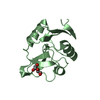
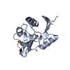
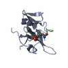
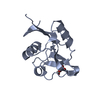
- PDB-4n38: Structure of langerin CRD I313 D288 complexed with GlcNAc-beta1-3... -
+ Open data

- Basic information
| Entry | Database: PDB / ID: 4n38 | |||||||||
|---|---|---|---|---|---|---|---|---|---|---|
| Title | Structure of langerin CRD I313 D288 complexed with GlcNAc-beta1-3Gal-beta1-4GlcNAc-beta-CH2CH2N3 | |||||||||
 Components Components | C-type lectin domain family 4 member K | |||||||||
 Keywords Keywords | SUGAR BINDING PROTEIN / CRD carbohydrate complex / carbohydrate-recognition domain / C-type lectin on Langerhans cells / Carbohydrate/Sugar Binding / receptor on Langerhans cells | |||||||||
| Function / homology |  Function and homology information Function and homology informationCross-presentation of soluble exogenous antigens (endosomes) / pattern recognition receptor activity / D-mannose binding / endocytic vesicle / clathrin-coated endocytic vesicle membrane / carbohydrate binding / early endosome membrane / defense response to virus / immune response / external side of plasma membrane / plasma membrane Similarity search - Function | |||||||||
| Biological species |  Homo sapiens (human) Homo sapiens (human) | |||||||||
| Method |  X-RAY DIFFRACTION / SYNCHROTRON / FOURIER SYNTHESIS / Resolution: 2 Å X-RAY DIFFRACTION / SYNCHROTRON / FOURIER SYNTHESIS / Resolution: 2 Å | |||||||||
 Authors Authors | Feinberg, H. / Rowntree, T.J.W. / Tan, S.L.W. / Drickamer, K. / Weis, W.I. / Taylor, M.E. | |||||||||
 Citation Citation | Journal: J.Biol.Chem. / Year: 2013 Title: Common polymorphisms in human langerin change specificity for glycan ligands. Authors: Feinberg, H. / Rowntree, T.J. / Tan, S.L. / Drickamer, K. / Weis, W.I. / Taylor, M.E. | |||||||||
| History |
|
- Structure visualization
| Structure viewer | Molecule: MolmilJmol/JSmol |
|---|
- Downloads & links
-Download
| PDBx/mmCIF format | 4n38.cif.gz | 130.2 KB | Display | PDBx/mmCIF format |
|---|---|---|---|---|
| PDB format | pdb4n38.ent.gz | 100.3 KB | Display | PDB format |
| PDBx/mmJSON format | 4n38.json.gz | Tree view | PDBx/mmJSON format | |
| Others |  Other downloads Other downloads |
-Validation report
| Arichive directory | https://data.pdbj.org/pub/pdb/validation_reports/n3/4n38ftp://data.pdbj.org/pub/pdb/validation_reports/n3/4n38 | HTTPS FTP |
|---|
-Related structure data
| Related structure data |  4n32C  4n33C  4n34C  4n35C  4n36C  4n37C  3p5gS C: citing same article ( S: Starting model for refinement |
|---|---|
| Similar structure data |


-Links
 PDBj
PDBj


- Assembly
| Deposited unit | 
| ||||||||
|---|---|---|---|---|---|---|---|---|---|
| 1 | 
| ||||||||
| 2 | 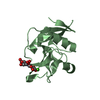
| ||||||||
| 3 | 
| ||||||||
| 4 | 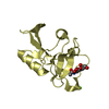
| ||||||||
| 5 | 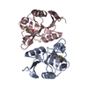
| ||||||||
| 6 | 
| ||||||||
| Unit cell |
| ||||||||
| Components on special symmetry positions |
|
-Components
| #1: Protein | Mass: 15562.339 Da / Num. of mol.: 4 / Fragment: carbohydrate recognition domain / Mutation: K313I, N288D Source method: isolated from a genetically manipulated source Source: (gene. exp.) Homo sapiens (human) / Gene: CD207, CLEC4K / Production host:  #2: Polysaccharide | 2-acetamido-2-deoxy-beta-D-glucopyranose-(1-3)-beta-D-galactopyranose Source method: isolated from a genetically manipulated source #3: Chemical | ChemComp-CA /   Mass: 40.078 Da / Num. of mol.: 4 / Source method: obtained synthetically / Formula: Ca Mass: 40.078 Da / Num. of mol.: 4 / Source method: obtained synthetically / Formula: Ca#4: Chemical | ChemComp-MG / |   Mass: 24.305 Da / Num. of mol.: 1 / Source method: obtained synthetically / Formula: Mg Mass: 24.305 Da / Num. of mol.: 1 / Source method: obtained synthetically / Formula: Mg#5: Water | ChemComp-HOH / |  Mass: 18.015 Da / Num. of mol.: 388 / Source method: isolated from a natural source / Formula: H2O Mass: 18.015 Da / Num. of mol.: 388 / Source method: isolated from a natural source / Formula: H2OHas protein modification | Y | |
|---|
-Experimental details
-Experiment
| Experiment | Method: X-RAY DIFFRACTION / Number of used crystals: 1 |
|---|
- Sample preparation
| Crystal | Density Matthews: 2.29 Å3/Da / Density % sol: 46.34 % |
|---|---|
| Crystal grow | Temperature: 295 K / Method: vapor diffusion, hanging drop / pH: 7 Details: Protein solution: 2.3-4.0 mg/mL langerin, 2.5 mM CaCl2, 10-100 mM Tris (pH 8.0), 25 mM NaCl and 15mM TR55. The reservoir solution contained 0.1 M HEPES (pH 7.0), 0.1-0.2 M MgCl2, and 20-35% ...Details: Protein solution: 2.3-4.0 mg/mL langerin, 2.5 mM CaCl2, 10-100 mM Tris (pH 8.0), 25 mM NaCl and 15mM TR55. The reservoir solution contained 0.1 M HEPES (pH 7.0), 0.1-0.2 M MgCl2, and 20-35% polyethylene glycol 3350, VAPOR DIFFUSION, HANGING DROP, temperature 295K |
-Data collection
| Diffraction | Mean temperature: 100 K | ||||||||||||||||||||||||||||||||||||||||||||||||||||||||||||||||||||||||||||||||||||||||
|---|---|---|---|---|---|---|---|---|---|---|---|---|---|---|---|---|---|---|---|---|---|---|---|---|---|---|---|---|---|---|---|---|---|---|---|---|---|---|---|---|---|---|---|---|---|---|---|---|---|---|---|---|---|---|---|---|---|---|---|---|---|---|---|---|---|---|---|---|---|---|---|---|---|---|---|---|---|---|---|---|---|---|---|---|---|---|---|---|---|
| Diffraction source | Source: SYNCHROTRON / Site: SSRL  / Beamline: BL11-1 / Wavelength: 0.97945 Å / Beamline: BL11-1 / Wavelength: 0.97945 Å | ||||||||||||||||||||||||||||||||||||||||||||||||||||||||||||||||||||||||||||||||||||||||
| Detector | Type: PSI PILATUS 6M / Detector: PIXEL / Date: May 7, 2012 | ||||||||||||||||||||||||||||||||||||||||||||||||||||||||||||||||||||||||||||||||||||||||
| Radiation | Monochromator: NA / Protocol: SINGLE WAVELENGTH / Monochromatic (M) / Laue (L): M / Scattering type: x-ray | ||||||||||||||||||||||||||||||||||||||||||||||||||||||||||||||||||||||||||||||||||||||||
| Radiation wavelength | Wavelength: 0.97945 Å / Relative weight: 1 | ||||||||||||||||||||||||||||||||||||||||||||||||||||||||||||||||||||||||||||||||||||||||
| Reflection | Resolution: 2→39.56 Å / Num. obs: 37595 / % possible obs: 99.1 % / Observed criterion σ(F): 0 / Observed criterion σ(I): 0 / Redundancy: 4.6 % / Biso Wilson estimate: 25.77 Å2 / Rsym value: 0.039 / Net I/σ(I): 25.1 | ||||||||||||||||||||||||||||||||||||||||||||||||||||||||||||||||||||||||||||||||||||||||
| Reflection shell | Diffraction-ID: 1
|
- Processing
| Software |
| ||||||||||||||||||||||||||||||||||||||||||||||||||||||||||||||||||||||||||||||||||||||||||||||||||
|---|---|---|---|---|---|---|---|---|---|---|---|---|---|---|---|---|---|---|---|---|---|---|---|---|---|---|---|---|---|---|---|---|---|---|---|---|---|---|---|---|---|---|---|---|---|---|---|---|---|---|---|---|---|---|---|---|---|---|---|---|---|---|---|---|---|---|---|---|---|---|---|---|---|---|---|---|---|---|---|---|---|---|---|---|---|---|---|---|---|---|---|---|---|---|---|---|---|---|---|
| Refinement | Method to determine structure: FOURIER SYNTHESIS Starting model: pdb entry 3P5G Resolution: 2→39.56 Å / Occupancy max: 1 / Occupancy min: 0.5 / FOM work R set: 0.8451 / SU ML: 0.26 / σ(F): 0 / Phase error: 23.08 / Stereochemistry target values: ML
| ||||||||||||||||||||||||||||||||||||||||||||||||||||||||||||||||||||||||||||||||||||||||||||||||||
| Solvent computation | Shrinkage radii: 0.98 Å / VDW probe radii: 1.2 Å / Solvent model: FLAT BULK SOLVENT MODEL / Bsol: 41.064 Å2 / ksol: 0.354 e/Å3 | ||||||||||||||||||||||||||||||||||||||||||||||||||||||||||||||||||||||||||||||||||||||||||||||||||
| Displacement parameters | Biso max: 84.96 Å2 / Biso mean: 28.1802 Å2 / Biso min: 10.84 Å2
| ||||||||||||||||||||||||||||||||||||||||||||||||||||||||||||||||||||||||||||||||||||||||||||||||||
| Refinement step | Cycle: LAST / Resolution: 2→39.56 Å
| ||||||||||||||||||||||||||||||||||||||||||||||||||||||||||||||||||||||||||||||||||||||||||||||||||
| Refine LS restraints |
| ||||||||||||||||||||||||||||||||||||||||||||||||||||||||||||||||||||||||||||||||||||||||||||||||||
| LS refinement shell | Refine-ID: X-RAY DIFFRACTION / Total num. of bins used: 13
|
