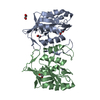
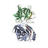
X: Histone-lysine N-methyltransferase, H3 lysine-9 specific SUVH5 A: Histone-lysine N-methyltransferase, H3 lysine-9 specific SUVH5 B: DNA (5'-D(*CP*TP*GP*AP*GP*GP*AP*GP*TP*AP*T)-3') C: DNA (5'-D(*TP*AP*CP*TP*(5CM)P*CP*TP*CP*AP*G)-3')
Histone-lysineN-methyltransferase, H3lysine-9specificSUVH5 / Histone H3-K9 methyltransferase 5 / H3-K9-HMTase 5 / Protein SET DOMAIN GROUP 9 / Suppressor of ...Histone H3-K9 methyltransferase 5 / H3-K9-HMTase 5 / Protein SET DOMAIN GROUP 9 / Suppressor of variegation 3-9 homolog protein 5 / Su(var)3-9 homolog protein 5
Mass: 18515.920 Da / Num. of mol.: 2 / Fragment: SUVH5 SRA Domain (UNP Residues 362-528) Source method: isolated from a genetically manipulated source Source: (gene. exp.) Arabidopsis thaliana (thale cress) / Gene: At2g35160, SDG9, SET9, SUVH5, T4C15.17 / Plasmid: pET SUMO / Production host: Escherichia coli (E. coli) / Strain (production host): Rosetta2 (DE3) References: UniProt: O82175, histone-lysine N-methyltransferase
#2: DNA chain
DNA (5'-D(*CP*TP*GP*AP*GP*GP*AP*GP*TP*AP*T)-3')
Mass: 3413.246 Da / Num. of mol.: 1 / Source method: obtained synthetically / Details: Chemically Synthesized
#3: DNA chain
DNA (5'-D(*TP*AP*CP*TP*(5CM)P*CP*TP*CP*AP*G)-3')
Mass: 2993.994 Da / Num. of mol.: 1 / Source method: obtained synthetically / Details: Chemically Synthesized
Resolution: 2.75→2.85 Å / Redundancy: 9.5 % / Rmerge(I) obs: 0.7 / Mean I/σ(I) obs: 2.2 / Rsym value: 0.59 / % possible all: 100
-
Processing
Software
Name
Version
Classification
ADSC
Quantum
datacollection
MOLREP
MR
phasing
PHENIX
(phenix.refine)
refinement
HKL-2000
datareduction
HKL-2000
datascaling
Refinement
Method to determine structure: MOLECULAR REPLACEMENT Starting model: SRA molecule from the SUVH5 SRA-fully methylated CG DNA crystallized in P42212 space group Resolution: 2.75→29.283 Å / SU ML: 1.09 / Cross valid method: THROUGHOUT / σ(F): 1.33 / Phase error: 25.07 / Stereochemistry target values: ML
Rfactor
Num. reflection
% reflection
Selection details
Rfree
0.2732
729
5.04 %
Random
Rwork
0.2294
-
-
-
obs
0.2315
14466
99.69 %
-
all
-
14500
-
-
Solvent computation
Shrinkage radii: 0.9 Å / VDW probe radii: 1.11 Å / Solvent model: FLAT BULK SOLVENT MODEL / Bsol: 53.6 Å2 / ksol: 0.29 e/Å3
Displacement parameters
Baniso -1
Baniso -2
Baniso -3
1-
7.7339 Å2
-0 Å2
-0 Å2
2-
-
7.7339 Å2
0 Å2
3-
-
-
-15.4678 Å2
Refinement step
Cycle: LAST / Resolution: 2.75→29.283 Å
Protein
Nucleic acid
Ligand
Solvent
Total
Num. atoms
2239
425
0
18
2682
Refine LS restraints
Refine-ID
Type
Dev ideal
Number
X-RAY DIFFRACTION
f_bond_d
0.007
2757
X-RAY DIFFRACTION
f_angle_d
1.294
3817
X-RAY DIFFRACTION
f_dihedral_angle_d
20.905
1040
X-RAY DIFFRACTION
f_chiral_restr
0.071
415
X-RAY DIFFRACTION
f_plane_restr
0.006
425
Refine LS restraints NCS
Ens-ID
Dom-ID
Auth asym-ID
Number
Refine-ID
Type
Rms dev position (Å)
1
1
X
300
X-RAY DIFFRACTION
POSITIONAL
1
2
A
300
X-RAY DIFFRACTION
POSITIONAL
0.026
2
1
X
64
X-RAY DIFFRACTION
POSITIONAL
2
2
A
64
X-RAY DIFFRACTION
POSITIONAL
0.03
3
1
X
645
X-RAY DIFFRACTION
POSITIONAL
3
2
A
645
X-RAY DIFFRACTION
POSITIONAL
0.041
LS refinement shell
Resolution (Å)
Rfactor Rfree
Num. reflection Rfree
Rfactor Rwork
Num. reflection Rwork
Refine-ID
% reflection obs (%)
2.75-2.9622
0.3353
149
0.2957
2655
X-RAY DIFFRACTION
100
2.9622-3.2599
0.3349
155
0.2555
2679
X-RAY DIFFRACTION
100
3.2599-3.7308
0.2384
125
0.2161
2734
X-RAY DIFFRACTION
100
3.7308-4.6973
0.232
152
0.1871
2763
X-RAY DIFFRACTION
100
4.6973-29.2842
0.2798
148
0.2351
2906
X-RAY DIFFRACTION
99
+
About Yorodumi
-
News
-
Feb 9, 2022. New format data for meta-information of EMDB entries
New format data for meta-information of EMDB entries
Version 3 of the EMDB header file is now the official format.
The previous official version 1.9 will be removed from the archive.
In the structure databanks used in Yorodumi, some data are registered as the other names, "COVID-19 virus" and "2019-nCoV". Here are the details of the virus and the list of structure data.
Jan 31, 2019. EMDB accession codes are about to change! (news from PDBe EMDB page)
EMDB accession codes are about to change! (news from PDBe EMDB page)
The allocation of 4 digits for EMDB accession codes will soon come to an end. Whilst these codes will remain in use, new EMDB accession codes will include an additional digit and will expand incrementally as the available range of codes is exhausted. The current 4-digit format prefixed with “EMD-” (i.e. EMD-XXXX) will advance to a 5-digit format (i.e. EMD-XXXXX), and so on. It is currently estimated that the 4-digit codes will be depleted around Spring 2019, at which point the 5-digit format will come into force.
The EM Navigator/Yorodumi systems omit the EMD- prefix.
Related info.:Q: What is EMD? / ID/Accession-code notation in Yorodumi/EM Navigator
Yorodumi is a browser for structure data from EMDB, PDB, SASBDB, etc.
This page is also the successor to EM Navigator detail page, and also detail information page/front-end page for Omokage search.
The word "yorodu" (or yorozu) is an old Japanese word meaning "ten thousand". "mi" (miru) is to see.
Related info.:EMDB / PDB / SASBDB / Comparison of 3 databanks / Yorodumi Search / Aug 31, 2016. New EM Navigator & Yorodumi / Yorodumi Papers / Jmol/JSmol / Function and homology information / Changes in new EM Navigator and Yorodumi
 Movie
Movie Controller


 Open data
 Basic information
 Components
 Keywords
 Function and homology information
 X-RAY DIFFRACTION /
  Authors
 Citation
 Structure visualization
 Downloads & links
 Other downloads


 PDBj
PDBj


 Assembly

 Mass: 18.015 Da / Num. of mol.: 18 / Source method: isolated from a natural source / Formula: H2O
 Sample preparation
 / Beamline: 24-ID-E / Wavelength: 0.97918 Å
 Processing