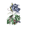
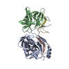
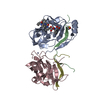
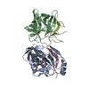
[English] 日本語
Yorodumi
- PDB-3kf2: The HCV NS3/NS4A protease apo structure -

+
Open data


ID or keywords:

Loading...

-
Basic information

Entry
Database: PDB / ID: 3kf2
TitleThe HCV NS3/NS4A protease apo structure
Components
  • 19-mer peptide from Genome polyprotein
  • Polyprotein
KeywordsHYDROLASE / HCV / NS3 / Protease / Apo
Function / homology
Function and homology information


symbiont-mediated suppression of host TRAF-mediated signal transduction => GO:0039527 / : / : / hepacivirin / host cell mitochondrial membrane / host cell lipid droplet / symbiont-mediated suppression of host TRAF-mediated signal transduction / transformation of host cell by virus / symbiont-mediated perturbation of host cell cycle G1/S transition checkpoint / host cell mitochondrion ...symbiont-mediated suppression of host TRAF-mediated signal transduction => GO:0039527 / : / : / hepacivirin / host cell mitochondrial membrane / host cell lipid droplet / symbiont-mediated suppression of host TRAF-mediated signal transduction / transformation of host cell by virus / symbiont-mediated perturbation of host cell cycle G1/S transition checkpoint / host cell mitochondrion / symbiont-mediated suppression of host JAK-STAT cascade via inhibition of STAT1 activity / symbiont-mediated suppression of host cytoplasmic pattern recognition receptor signaling pathway via inhibition of MAVS activity / ribonucleoside triphosphate phosphatase activity / lipid droplet / viral capsid / nucleoside-triphosphate phosphatase / protein complex oligomerization / monoatomic ion channel activity / viral nucleocapsid / clathrin-dependent endocytosis of virus by host cell / membrane => GO:0016020 / RNA helicase activity / host cell perinuclear region of cytoplasm / host cell endoplasmic reticulum membrane / symbiont-mediated suppression of host type I interferon-mediated signaling pathway / RNA helicase / ribonucleoprotein complex / symbiont entry into host cell / induction by virus of host autophagy / RNA-directed RNA polymerase / viral RNA genome replication / cysteine-type endopeptidase activity / serine-type endopeptidase activity / RNA-dependent RNA polymerase activity / fusion of virus membrane with host endosome membrane / viral envelope / host cell nucleus / virion attachment to host cell / endoplasmic reticulum membrane / host cell plasma membrane / structural molecule activity / virion membrane / proteolysis / RNA binding / zinc ion binding / extracellular region / ATP binding / nucleus / plasma membrane / cytoplasm
Similarity search - Function
Thrombin, subunit H - #120 / Hepatitis C virus core protein, chain A superfamily / Hepatitus C virus, Non-structural 5a protein, C-terminal / Hepatitis C virus NS5A, 1B domain superfamily / Hepatitis C virus non-structural protein NS2, C-terminal domain / Hepatitis C virus non-structural protein NS2, N-terminal domain / Hepatitis C virus non-structural protein NS2 / HCV NS5a protein C-terminal region / Hepatitis C virus, Non-structural protein NS4b / Hepatitis C virus, Core protein, N-terminal ...Thrombin, subunit H - #120 / Hepatitis C virus core protein, chain A superfamily / Hepatitus C virus, Non-structural 5a protein, C-terminal / Hepatitis C virus NS5A, 1B domain superfamily / Hepatitis C virus non-structural protein NS2, C-terminal domain / Hepatitis C virus non-structural protein NS2, N-terminal domain / Hepatitis C virus non-structural protein NS2 / HCV NS5a protein C-terminal region / Hepatitis C virus, Non-structural protein NS4b / Hepatitis C virus, Core protein, N-terminal / Hepatitis C virus non-structural protein NS4b / Hepatitis C virus capsid protein / Hepatitis C virus, Non-structural protein NS2 / Hepatitis C virus, Non-structural 5a protein / Hepatitis C virus, Non-structural 5a protein, domain 1a / Hepatitis C virus non-structural 5a, 1B domain / NS5A domain 1a superfamily / Hepatitis C virus non-structural 5a protein membrane anchor / Hepatitis C virus non-structural 5a zinc finger domain / Hepatitis C virus non-structural 5a domain 1b / Hepacivirus nonstructural protein 2 (NS2) protease domain profile. / Hepatitis C virus, Non-structural protein NS4a / Hepatitis C virus non-structural protein NS4a / Hepatitis C virus, Core protein, C-terminal / Hepatitis C virus core protein / Hepatitis C virus, Non-structural protein E2/NS1 / Hepatitis C virus non-structural protein E2/NS1 / Hepatitis C virus, Envelope glycoprotein E1 / Hepatitis C virus envelope glycoprotein E1 / RNA dependent RNA polymerase, hepatitis C virus / Viral RNA dependent RNA polymerase / Hepatitis C virus, NS3 protease, Peptidase S29 / Hepatitis C virus NS3 protease / Hepacivirus/Pegivirus NS3 protease domain profile. / DEAD box, Flavivirus / Flavivirus DEAD domain / helicase superfamily c-terminal domain / Trypsin-like serine proteases / Superfamilies 1 and 2 helicase C-terminal domain profile. / Superfamilies 1 and 2 helicase ATP-binding type-1 domain profile. / DEAD-like helicases superfamily / Helicase, C-terminal / Helicase superfamily 1/2, ATP-binding domain / Thrombin, subunit H / Reverse transcriptase/Diguanylate cyclase domain / RNA-directed RNA polymerase, catalytic domain / RdRp of positive ssRNA viruses catalytic domain profile. / Peptidase S1, PA clan, chymotrypsin-like fold / Peptidase S1, PA clan / DNA/RNA polymerase superfamily / Beta Barrel / P-loop containing nucleoside triphosphate hydrolase / Mainly Beta
Similarity search - Domain/homology
Genome polyprotein / Capsid protein C / Genome polyprotein
Similarity search - Component
Biological speciesHepatitis C virus
MethodX-RAY DIFFRACTION / SYNCHROTRON / MOLECULAR REPLACEMENT / molecular replacement / Resolution: 2.5 Å
AuthorsLindberg, J.D. / Nystrom, S. / Cummings, M.D.
CitationJournal: Angew.Chem.Int.Ed.Engl. / Year: 2010
Title: Induced-Fit Binding of the Macrocyclic Noncovalent Inhibitor TMC435 to its HCV NS3/NS4A Protease Target
Authors: Cummings, M.D. / Lindberg, J.D. / Lin, T.-I. / de Kock, H. / Lenz, O. / Lilja, E. / Fellander, S. / Baraznenok, V. / Nystrom, S. / Nilsson, M. / Vrang, L. / Edlund, M. / Rosenquist, A. / ...Authors: Cummings, M.D. / Lindberg, J.D. / Lin, T.-I. / de Kock, H. / Lenz, O. / Lilja, E. / Fellander, S. / Baraznenok, V. / Nystrom, S. / Nilsson, M. / Vrang, L. / Edlund, M. / Rosenquist, A. / Samuelsson, B. / Raboisson, P. / Simmen, K.
History
DepositionOct 27, 2009Deposition site: RCSB / Processing site: PDBJ
Revision 1.0Mar 9, 2010Provider: repository / Type: Initial release
Revision 1.1Jul 13, 2011Group: Version format compliance
Revision 1.2Nov 10, 2021Group: Database references / Derived calculations
Category: database_2 / struct_conn ...database_2 / struct_conn / struct_ref_seq_dif / struct_site
Item: _database_2.pdbx_DOI / _database_2.pdbx_database_accession ..._database_2.pdbx_DOI / _database_2.pdbx_database_accession / _struct_conn.pdbx_dist_value / _struct_conn.ptnr1_auth_asym_id / _struct_conn.ptnr1_auth_comp_id / _struct_conn.ptnr1_auth_seq_id / _struct_conn.ptnr1_label_asym_id / _struct_conn.ptnr1_label_atom_id / _struct_conn.ptnr1_label_comp_id / _struct_conn.ptnr1_label_seq_id / _struct_conn.ptnr2_auth_asym_id / _struct_conn.ptnr2_auth_comp_id / _struct_conn.ptnr2_auth_seq_id / _struct_conn.ptnr2_label_asym_id / _struct_conn.ptnr2_label_atom_id / _struct_conn.ptnr2_label_comp_id / _struct_ref_seq_dif.details / _struct_site.pdbx_auth_asym_id / _struct_site.pdbx_auth_comp_id / _struct_site.pdbx_auth_seq_id
Revision 1.3Nov 1, 2023Group: Data collection / Refinement description
Category: chem_comp_atom / chem_comp_bond / pdbx_initial_refinement_model

-
Structure visualization

Structure viewerMolecule:
MolmilJmol/JSmol

Downloads & links

-
Assembly

Deposited unit
A: Polyprotein
C: 19-mer peptide from Genome polyprotein
B: Polyprotein
D: 19-mer peptide from Genome polyprotein
hetero molecules


Theoretical massNumber of molelcules
Total (without water)47,3856
Polymers47,2554
Non-polymers1312
Water72140
1


  • Idetical with deposited unit
  • defined by author&software
TypeNameSymmetry operationNumber
identity operation1_555x,y,z1
Buried area6690 Å2
ΔGint-120 kcal/mol
Surface area16130 Å2
MethodPISA
Unit cell
Length a, b, c (Å)224.924, 224.924, 75.545
Angle α, β, γ (deg.)90.000, 90.000, 120.000
Int Tables number155
Space group name H-MH32

-
Components

#1: Protein Polyprotein


Mass: 21233.225 Da / Num. of mol.: 2 / Fragment: NS3 protease domain, UNP residues 149-329 / Mutation: G176E
Source method: isolated from a genetically manipulated source
Source: (gene. exp.) Hepatitis C virus / Production host: Escherichia coli (E. coli)
References: UniProt: B1PBR5, UniProt: B2Y2M9*PLUS, hepacivirin
#2: Protein/peptide 19-mer peptide from Genome polyprotein


Mass: 2394.039 Da / Num. of mol.: 2 / Fragment: NS4a peptide, UNP residues 1682-1700 / Source method: obtained synthetically / Details: chemical synthesis / Source: (synth.) Hepatitis C virus / References: UniProt: Q6GYR8
#3: Chemical ChemComp-ZN / ZINC ION


Mass: 65.409 Da / Num. of mol.: 2 / Source method: obtained synthetically / Formula: Zn
#4: Water ChemComp-HOH / water


Mass: 18.015 Da / Num. of mol.: 40 / Source method: isolated from a natural source / Formula: H2O

-
Experimental details

-
Experiment

ExperimentMethod: X-RAY DIFFRACTION / Number of used crystals: 1

-
Sample preparation

CrystalDensity Matthews: 3.89 Å3/Da / Density % sol: 68.39 % / Mosaicity: 0.323 °
Crystal growTemperature: 277 K / Method: vapor diffusion, hanging drop / pH: 6.2
Details: 1.2M NaCl, 50mM MES, 50mM Na/K Phosphate, 10mM BME, pH 6.2, VAPOR DIFFUSION, HANGING DROP, temperature 277K

-
Data collection

DiffractionMean temperature: 100 K
Diffraction sourceSource: SYNCHROTRON / Site: ESRF / Beamline: ID23-1 / Wavelength: 0.98 Å
DetectorType: ADSC QUANTUM 1 / Detector: CCD / Date: Jan 30, 2008
RadiationMonochromator: GRAPHITE / Protocol: SINGLE WAVELENGTH / Monochromatic (M) / Laue (L): M / Scattering type: x-ray
Radiation wavelengthWavelength: 0.98 Å / Relative weight: 1
ReflectionResolution: 2.5→112.509 Å / Num. obs: 25152 / % possible obs: 99.6 % / Observed criterion σ(F): 2 / Observed criterion σ(I): 2 / Redundancy: 4.9 % / Rmerge(I) obs: 0.111 / Rsym value: 0.111 / Net I/σ(I): 12.8 / Num. measured all: 124042
Reflection shellResolution: 2.5→2.64 Å / Redundancy: 5 % / Rmerge(I) obs: 0.627 / Mean I/σ(I) obs: 1.1 / Num. measured all: 18310 / Num. unique all: 3664 / Rsym value: 0.627 / % possible all: 100

-
Phasing

PhasingMethod: molecular replacement
Phasing MRModel details: Phaser MODE: MR_AUTO
Highest resolutionLowest resolution
Rotation2.5 Å112.46 Å
Translation2.5 Å112.46 Å

-
Processing

Software
NameVersionClassificationNB
MOSFLMdata reduction
SCALA3.2.25data scaling
PHASER1.3.3phasing
REFMACrefinement
PDB_EXTRACT3.005data extraction
DNAdata collection
RefinementMethod to determine structure: MOLECULAR REPLACEMENT
Starting model: PDB ENTRY 2A4G
Resolution: 2.5→112.46 Å / Cor.coef. Fo:Fc: 0.935 / Cor.coef. Fo:Fc free: 0.911 / WRfactor Rfree: 0.227 / WRfactor Rwork: 0.199 / Occupancy max: 1 / Occupancy min: 1 / FOM work R set: 0.858 / SU B: 6.2 / SU ML: 0.138 / SU R Cruickshank DPI: 0.245 / SU Rfree: 0.205 / Cross valid method: THROUGHOUT / σ(F): 0 / ESU R: 0.245 / ESU R Free: 0.205 / Stereochemistry target values: MAXIMUM LIKELIHOOD / Details: HYDROGENS HAVE BEEN ADDED IN THE RIDING POSITIONS
RfactorNum. reflection% reflectionSelection details
Rfree0.235 1285 5.1 %RANDOM
Rwork0.205 ---
all0.217 25286 --
obs0.207 25152 99.47 %-
Solvent computationIon probe radii: 0.8 Å / Shrinkage radii: 0.8 Å / VDW probe radii: 1.2 Å / Solvent model: MASK
Displacement parametersBiso max: 74.55 Å2 / Biso mean: 33.813 Å2 / Biso min: 8.51 Å2
Baniso -1Baniso -2Baniso -3
1--0.11 Å2-0.06 Å20 Å2
2---0.11 Å20 Å2
3---0.17 Å2
Refinement stepCycle: LAST / Resolution: 2.5→112.46 Å
ProteinNucleic acidLigandSolventTotal
Num. atoms2740 0 2 40 2782
Refine LS restraints
Refine-IDTypeDev idealDev ideal targetNumber
X-RAY DIFFRACTIONr_bond_refined_d0.0120.0222793
X-RAY DIFFRACTIONr_angle_refined_deg1.5651.9753805
X-RAY DIFFRACTIONr_dihedral_angle_1_deg6.8155368
X-RAY DIFFRACTIONr_dihedral_angle_2_deg30.74621.7293
X-RAY DIFFRACTIONr_dihedral_angle_3_deg20.18815443
X-RAY DIFFRACTIONr_dihedral_angle_4_deg19.6551526
X-RAY DIFFRACTIONr_chiral_restr0.1070.2455
X-RAY DIFFRACTIONr_gen_planes_refined0.0050.022072
X-RAY DIFFRACTIONr_nbd_refined0.2210.21127
X-RAY DIFFRACTIONr_nbtor_refined0.3130.21901
X-RAY DIFFRACTIONr_xyhbond_nbd_refined0.1480.2117
X-RAY DIFFRACTIONr_symmetry_vdw_refined0.1740.236
X-RAY DIFFRACTIONr_symmetry_hbond_refined0.1290.22
X-RAY DIFFRACTIONr_mcbond_it0.8241.51879
X-RAY DIFFRACTIONr_mcangle_it1.48522989
X-RAY DIFFRACTIONr_scbond_it2.06431014
X-RAY DIFFRACTIONr_scangle_it3.574.5816
LS refinement shellResolution: 2.5→2.565 Å / Total num. of bins used: 20
RfactorNum. reflection% reflection
Rfree0.361 90 -
Rwork0.324 1737 -
all-1827 -
obs--99.95 %

+
About Yorodumi

-
News

-
Feb 9, 2022. New format data for meta-information of EMDB entries

New format data for meta-information of EMDB entries

  • Version 3 of the EMDB header file is now the official format.
  • The previous official version 1.9 will be removed from the archive.

Related info.:EMDB header

External links:wwPDB to switch to version 3 of the EMDB data model

-
Aug 12, 2020. Covid-19 info

Covid-19 info

URL: https://pdbjlvh1.pdbj.org/emnavi/covid19.php

New page: Covid-19 featured information page in EM Navigator.

Related info.:Covid-19 info / Mar 5, 2020. Novel coronavirus structure data

+
Mar 5, 2020. Novel coronavirus structure data

Novel coronavirus structure data

Related info.:Yorodumi Speices / Aug 12, 2020. Covid-19 info

External links:COVID-19 featured content - PDBj / Molecule of the Month (242):Coronavirus Proteases

+
Jan 31, 2019. EMDB accession codes are about to change! (news from PDBe EMDB page)

EMDB accession codes are about to change! (news from PDBe EMDB page)

  • The allocation of 4 digits for EMDB accession codes will soon come to an end. Whilst these codes will remain in use, new EMDB accession codes will include an additional digit and will expand incrementally as the available range of codes is exhausted. The current 4-digit format prefixed with “EMD-” (i.e. EMD-XXXX) will advance to a 5-digit format (i.e. EMD-XXXXX), and so on. It is currently estimated that the 4-digit codes will be depleted around Spring 2019, at which point the 5-digit format will come into force.
  • The EM Navigator/Yorodumi systems omit the EMD- prefix.

Related info.:Q: What is EMD? / ID/Accession-code notation in Yorodumi/EM Navigator

External links:EMDB Accession Codes are Changing Soon! / Contact to PDBj

+
Jul 12, 2017. Major update of PDB

Major update of PDB

  • wwPDB released updated PDB data conforming to the new PDBx/mmCIF dictionary.
  • This is a major update changing the version number from 4 to 5, and with Remediation, in which all the entries are updated.
  • In this update, many items about electron microscopy experimental information are reorganized (e.g. em_software).
  • Now, EM Navigator and Yorodumi are based on the updated data.

External links:wwPDB Remediation / Enriched Model Files Conforming to OneDep Data Standards Now Available in the PDB FTP Archive

-
Yorodumi

Thousand views of thousand structures

  • Yorodumi is a browser for structure data from EMDB, PDB, SASBDB, etc.
  • This page is also the successor to EM Navigator detail page, and also detail information page/front-end page for Omokage search.
  • The word "yorodu" (or yorozu) is an old Japanese word meaning "ten thousand". "mi" (miru) is to see.

Related info.:EMDB / PDB / SASBDB / Comparison of 3 databanks / Yorodumi Search / Aug 31, 2016. New EM Navigator & Yorodumi / Yorodumi Papers / Jmol/JSmol / Function and homology information / Changes in new EM Navigator and Yorodumi

Read more