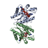
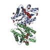
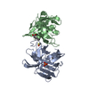
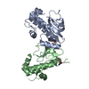
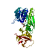
Histone-lysineN-methyltransferase, H3lysine-9specificSUVH5 / Histone H3-K9 methyltransferase 5 / H3-K9-HMTase 5 / Protein SET DOMAIN GROUP 9 / Suppressor of ...Histone H3-K9 methyltransferase 5 / H3-K9-HMTase 5 / Protein SET DOMAIN GROUP 9 / Suppressor of variegation 3-9 homolog protein 5 / Su(var)3-9 homolog protein 5
Mass: 18515.920 Da / Num. of mol.: 2 / Fragment: SUVH5 SRA Domain (UNP Residues 362-528) Source method: isolated from a genetically manipulated source Details: Cleavable N-terminal His-SUmo tag / Source: (gene. exp.) Arabidopsis thaliana (thale cress) / Gene: At2g35160, SDG9, SET9, SUVH5, T4C15.17 / Plasmid: pET SUMO / Production host: Escherichia coli (E. coli) / Strain (production host): Rosetta2 (DE3) References: UniProt: O82175, histone-lysine N-methyltransferase
#2: DNA chain
DNA (5'-D(*TP*CP*CP*AP*(5CM)P*GP*TP*CP*AP*G)-3')
Mass: 3019.008 Da / Num. of mol.: 1 / Source method: obtained synthetically / Details: Chemically synthesized
#3: DNA chain
DNA (5'-D(*CP*TP*GP*AP*CP*GP*TP*GP*GP*A)-3')
Mass: 3085.028 Da / Num. of mol.: 1 / Source method: obtained synthetically / Details: Chemically synthesized
Mass: 18.015 Da / Num. of mol.: 51 / Source method: isolated from a natural source / Formula: H2O
-
Experimental details
-
Experiment
Experiment
Method: X-RAY DIFFRACTION / Number of used crystals: 1
-
Sample preparation
Crystal
Density Matthews: 2.51 Å3/Da / Density % sol: 51.01 %
Crystal grow
Temperature: 291 K / Method: vapor diffusion, sitting drop / pH: 6.5 Details: 19% PEG4000, 100 mM MES, pH 6.5, 200 mM NaCl and 3 mM spermine, VAPOR DIFFUSION, SITTING DROP, temperature 291K
Method to determine structure: MOLECULAR REPLACEMENT Starting model: SRA Molecule from the SUVH5 SRA-fully methylated Cg DNA crystallized in space group P42212 Resolution: 2.3704→19.095 Å / SU ML: 0.35 / σ(F): 1.39 / Phase error: 27.86 / Stereochemistry target values: ML
Rfactor
Num. reflection
% reflection
Rfree
0.2628
873
5.07 %
Rwork
0.2246
-
-
obs
0.2265
17221
98.78 %
Solvent computation
Shrinkage radii: 0.83 Å / VDW probe radii: 1.1 Å / Solvent model: FLAT BULK SOLVENT MODEL / Bsol: 51.199 Å2 / ksol: 0.306 e/Å3
Displacement parameters
Baniso -1
Baniso -2
Baniso -3
1-
1.8426 Å2
-0 Å2
0 Å2
2-
-
1.8426 Å2
0 Å2
3-
-
-
-7.7466 Å2
Refinement step
Cycle: LAST / Resolution: 2.3704→19.095 Å
Protein
Nucleic acid
Ligand
Solvent
Total
Num. atoms
2173
405
1
51
2630
Refine LS restraints
Refine-ID
Type
Dev ideal
Number
X-RAY DIFFRACTION
f_bond_d
0.009
3114
X-RAY DIFFRACTION
f_angle_d
1.378
4382
X-RAY DIFFRACTION
f_dihedral_angle_d
22.458
1195
X-RAY DIFFRACTION
f_chiral_restr
0.082
486
X-RAY DIFFRACTION
f_plane_restr
0.007
433
Refine LS restraints NCS
Ens-ID
Dom-ID
Auth asym-ID
Number
Refine-ID
Type
Rms dev position (Å)
1
1
X
966
X-RAY DIFFRACTION
POSITIONAL
1
2
A
966
X-RAY DIFFRACTION
POSITIONAL
0.031
LS refinement shell
Resolution (Å)
Rfactor Rfree
Num. reflection Rfree
Rfactor Rwork
Num. reflection Rwork
Refine-ID
% reflection obs (%)
2.3704-2.5186
0.3309
135
0.3062
2584
X-RAY DIFFRACTION
94
2.5186-2.7125
0.3171
132
0.2631
2737
X-RAY DIFFRACTION
100
2.7125-2.9846
0.3162
146
0.276
2758
X-RAY DIFFRACTION
100
2.9846-3.4143
0.3028
142
0.2531
2751
X-RAY DIFFRACTION
100
3.4143-4.2936
0.2668
170
0.2141
2738
X-RAY DIFFRACTION
100
4.2936-19.0954
0.2124
148
0.1933
2780
X-RAY DIFFRACTION
99
+
About Yorodumi
-
News
-
Feb 9, 2022. New format data for meta-information of EMDB entries
New format data for meta-information of EMDB entries
Version 3 of the EMDB header file is now the official format.
The previous official version 1.9 will be removed from the archive.
In the structure databanks used in Yorodumi, some data are registered as the other names, "COVID-19 virus" and "2019-nCoV". Here are the details of the virus and the list of structure data.
Jan 31, 2019. EMDB accession codes are about to change! (news from PDBe EMDB page)
EMDB accession codes are about to change! (news from PDBe EMDB page)
The allocation of 4 digits for EMDB accession codes will soon come to an end. Whilst these codes will remain in use, new EMDB accession codes will include an additional digit and will expand incrementally as the available range of codes is exhausted. The current 4-digit format prefixed with “EMD-” (i.e. EMD-XXXX) will advance to a 5-digit format (i.e. EMD-XXXXX), and so on. It is currently estimated that the 4-digit codes will be depleted around Spring 2019, at which point the 5-digit format will come into force.
The EM Navigator/Yorodumi systems omit the EMD- prefix.
Related info.:Q: What is EMD? / ID/Accession-code notation in Yorodumi/EM Navigator
Yorodumi is a browser for structure data from EMDB, PDB, SASBDB, etc.
This page is also the successor to EM Navigator detail page, and also detail information page/front-end page for Omokage search.
The word "yorodu" (or yorozu) is an old Japanese word meaning "ten thousand". "mi" (miru) is to see.
Related info.:EMDB / PDB / SASBDB / Comparison of 3 databanks / Yorodumi Search / Aug 31, 2016. New EM Navigator & Yorodumi / Yorodumi Papers / Jmol/JSmol / Function and homology information / Changes in new EM Navigator and Yorodumi
 Movie
Movie Controller


 Open data
 Basic information
 Components
 Keywords
 Function and homology information
 X-RAY DIFFRACTION /
  Authors
 Citation
 Structure visualization
 Downloads & links
 Other downloads


 PDBj
PDBj
 Assembly
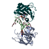


 Mass: 35.453 Da / Num. of mol.: 1 / Source method: obtained synthetically / Formula: Cl
 Mass: 18.015 Da / Num. of mol.: 51 / Source method: isolated from a natural source / Formula: H2O
 Sample preparation
 / Beamline: 24-ID-C / Wavelength: 0.97918 Å
 Processing