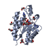
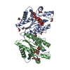
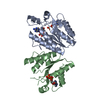
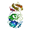
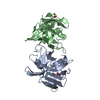
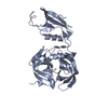
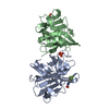
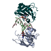
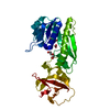
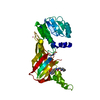
Entry Database : PDB / ID : 4bzqTitle Structure of the Mycobacterium tuberculosis APS kinase CysC in complex with ADP and APS BIFUNCTIONAL ENZYME CYSN/CYSC Keywords / / Function / homology Function Domain/homology Component
/ / / / / / / / / / / / / / / / / / / / / / / / / / / / / / / / / / / / / / / / / / / / / / / / / / / / / / / / / / Biological species MYCOBACTERIUM TUBERCULOSIS (bacteria)Method / / / Resolution : 2.1 Å Authors Poyraz, O. / Schnell, R. / Schneider, G. Journal : Plos One / Year : 2015Title : Crystal Structures of the Kinase Domain of the Sulfate-Activating Complex in Mycobacterium Tuberculosis.Authors : Poyraz, O. / Brunner, K. / Lohkamp, B. / Axelsson, H. / Hammarstrom, L.G.J. / Schnell, R. / Schneider, G. History Deposition Jul 29, 2013 Deposition site / Processing site Revision 1.0 Aug 13, 2014 Provider / Type Revision 1.1 Apr 8, 2015 Group Revision 1.2 May 16, 2018 Group / Category / Item Revision 1.3 Dec 20, 2023 Group Data collection / Database references ... Data collection / Database references / Derived calculations / Other / Refinement description Category chem_comp_atom / chem_comp_bond ... chem_comp_atom / chem_comp_bond / database_2 / pdbx_database_status / pdbx_initial_refinement_model / struct_ncs_dom_lim / struct_site Item _database_2.pdbx_DOI / _database_2.pdbx_database_accession ... _database_2.pdbx_DOI / _database_2.pdbx_database_accession / _pdbx_database_status.status_code_sf / _struct_ncs_dom_lim.beg_auth_comp_id / _struct_ncs_dom_lim.beg_label_asym_id / _struct_ncs_dom_lim.beg_label_comp_id / _struct_ncs_dom_lim.beg_label_seq_id / _struct_ncs_dom_lim.end_auth_comp_id / _struct_ncs_dom_lim.end_label_asym_id / _struct_ncs_dom_lim.end_label_comp_id / _struct_ncs_dom_lim.end_label_seq_id / _struct_site.pdbx_auth_asym_id / _struct_site.pdbx_auth_comp_id / _struct_site.pdbx_auth_seq_id
Show all Show less
 Movie
Movie Controller

 Yorodumi
 Open data
 Basic information
 Components
 Keywords
 Function and homology information
 MYCOBACTERIUM TUBERCULOSIS (bacteria)
 X-RAY DIFFRACTION /
  Authors
 Citation
 Structure visualization
 Downloads & links
 Other downloads
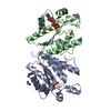


 PDBj
PDBj


 Assembly
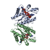


 Type: RNA linking / Mass: 427.284 Da / Num. of mol.: 2 / Source method: obtained synthetically / Formula: C10H14N5O10PS
 Mass: 427.201 Da / Num. of mol.: 2 / Source method: obtained synthetically / Formula: C10H15N5O10P2 / Comment: ADP, energy-carrying molecule*YM
 Mass: 192.124 Da / Num. of mol.: 2 / Source method: obtained synthetically / Formula: C6H8O7
 Mass: 62.068 Da / Num. of mol.: 2 / Source method: obtained synthetically / Formula: C2H6O2
 Sample preparation
 / Beamline: BM14 / Type:
  Processing