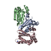
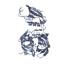
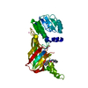
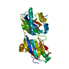
TRANSFERASE / NMP-kinase fold / protein in complex with nucleic acid
Function / homology
Function and homology information
3'-phosphoadenosine 5'-phosphosulfate biosynthetic process / Transport and metabolism of PAPS / sulfate adenylyltransferase / adenylyl-sulfate kinase / adenylylsulfate kinase activity / sulfate adenylyltransferase (ATP) activity / Metabolism of ingested H2SeO4 and H2SeO3 into H2Se / sulfate assimilation / nucleotidyltransferase activity / skeletal system development ...3'-phosphoadenosine 5'-phosphosulfate biosynthetic process / Transport and metabolism of PAPS / sulfate adenylyltransferase / adenylyl-sulfate kinase / adenylylsulfate kinase activity / sulfate adenylyltransferase (ATP) activity / Metabolism of ingested H2SeO4 and H2SeO3 into H2Se / sulfate assimilation / nucleotidyltransferase activity / skeletal system development / Signaling by BRAF and RAF1 fusions / protein homodimerization activity / ATP binding / nucleus / cytosol Similarity search - Function
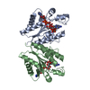
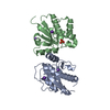
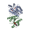
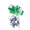
Journal: J.Biol.Chem. / Year: 2007 Title: Structural mechanism for substrate inhibition of the adenosine 5'-phosphosulfate kinase domain of human 3'-phosphoadenosine 5'-phosphosulfate synthetase 1 and its ramifications for enzyme regulation. Authors: Sekulic, N. / Konrad, M. / Lavie, A.
Mass: 19874.469 Da / Num. of mol.: 2 / Fragment: APS-kinase domain (residues 51-226) Source method: isolated from a genetically manipulated source Source: (gene. exp.) Homo sapiens (human) / Gene: PAPSS1, ATPSK1, PAPSS Plasmid: PGEX-RB in which the thrombin clevage site was replaced by TEV site Production host: Escherichia coli (E. coli) / Strain (production host): BL-21 Codon plus / References: UniProt: O43252
Resolution: 1.4→20 Å / Cor.coef. Fo:Fc: 0.952 / Cor.coef. Fo:Fc free: 0.945 / SU B: 1.037 / SU ML: 0.042 / Cross valid method: THROUGHOUT / ESU R: 0.076 / ESU R Free: 0.076 / Stereochemistry target values: MAXIMUM LIKELIHOOD / Details: HYDROGENS HAVE BEEN ADDED IN THE RIDING POSITIONS
Rfactor
Num. reflection
% reflection
Selection details
Rfree
0.21981
6493
10.1 %
RANDOM
Rwork
0.19433
-
-
-
obs
0.19695
57725
87.89 %
-
Solvent computation
Ion probe radii: 0.8 Å / Shrinkage radii: 0.8 Å / VDW probe radii: 1.4 Å / Solvent model: MASK
Displacement parameters
Biso mean: 16.825 Å2
Baniso -1
Baniso -2
Baniso -3
1-
1.02 Å2
0 Å2
0 Å2
2-
-
-0.16 Å2
0 Å2
3-
-
-
-0.87 Å2
Refinement step
Cycle: LAST / Resolution: 1.4→20 Å
Protein
Nucleic acid
Ligand
Solvent
Total
Num. atoms
2600
0
82
331
3013
Refine LS restraints
Refine-ID
Type
Dev ideal
Dev ideal target
Number
X-RAY DIFFRACTION
r_bond_refined_d
0.013
0.022
2750
X-RAY DIFFRACTION
r_angle_refined_deg
1.937
2.007
3743
X-RAY DIFFRACTION
r_dihedral_angle_1_deg
5.351
5
334
X-RAY DIFFRACTION
r_dihedral_angle_2_deg
29.554
24.884
129
X-RAY DIFFRACTION
r_dihedral_angle_3_deg
12.651
15
457
X-RAY DIFFRACTION
r_dihedral_angle_4_deg
14.493
15
18
X-RAY DIFFRACTION
r_chiral_restr
0.099
0.2
420
X-RAY DIFFRACTION
r_gen_planes_refined
0.007
0.02
2061
X-RAY DIFFRACTION
r_nbd_refined
0.215
0.2
1344
X-RAY DIFFRACTION
r_nbtor_refined
0.314
0.2
1888
X-RAY DIFFRACTION
r_xyhbond_nbd_refined
0.237
0.2
302
X-RAY DIFFRACTION
r_symmetry_vdw_refined
0.183
0.2
42
X-RAY DIFFRACTION
r_symmetry_hbond_refined
0.16
0.2
21
X-RAY DIFFRACTION
r_mcbond_it
1.131
1.5
1711
X-RAY DIFFRACTION
r_mcangle_it
1.65
2
2695
X-RAY DIFFRACTION
r_scbond_it
2.644
3
1149
X-RAY DIFFRACTION
r_scangle_it
3.965
4.5
1048
LS refinement shell
Resolution: 1.4→1.435 Å / Total num. of bins used: 20
Rfactor
Num. reflection
% reflection
Rfree
0.302
427
-
Rwork
0.255
3935
-
obs
-
-
81.95 %
+
About Yorodumi
-
News
-
Feb 9, 2022. New format data for meta-information of EMDB entries
New format data for meta-information of EMDB entries
Version 3 of the EMDB header file is now the official format.
The previous official version 1.9 will be removed from the archive.
In the structure databanks used in Yorodumi, some data are registered as the other names, "COVID-19 virus" and "2019-nCoV". Here are the details of the virus and the list of structure data.
Jan 31, 2019. EMDB accession codes are about to change! (news from PDBe EMDB page)
EMDB accession codes are about to change! (news from PDBe EMDB page)
The allocation of 4 digits for EMDB accession codes will soon come to an end. Whilst these codes will remain in use, new EMDB accession codes will include an additional digit and will expand incrementally as the available range of codes is exhausted. The current 4-digit format prefixed with “EMD-” (i.e. EMD-XXXX) will advance to a 5-digit format (i.e. EMD-XXXXX), and so on. It is currently estimated that the 4-digit codes will be depleted around Spring 2019, at which point the 5-digit format will come into force.
The EM Navigator/Yorodumi systems omit the EMD- prefix.
Related info.:Q: What is EMD? / ID/Accession-code notation in Yorodumi/EM Navigator
Yorodumi is a browser for structure data from EMDB, PDB, SASBDB, etc.
This page is also the successor to EM Navigator detail page, and also detail information page/front-end page for Omokage search.
The word "yorodu" (or yorozu) is an old Japanese word meaning "ten thousand". "mi" (miru) is to see.
Related info.:EMDB / PDB / SASBDB / Comparison of 3 databanks / Yorodumi Search / Aug 31, 2016. New EM Navigator & Yorodumi / Yorodumi Papers / Jmol/JSmol / Function and homology information / Changes in new EM Navigator and Yorodumi
 Movie
Movie Controller

 Yorodumi
 Open data
 Basic information
 Components
 Keywords
 Function and homology information
 Homo sapiens (human)
 X-RAY DIFFRACTION /
  Authors
 Citation
 Structure visualization
 Downloads & links
 Other downloads


 PDBj
PDBj
 Assembly
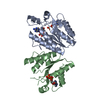

 Mass: 489.249 Da / Num. of mol.: 1 / Source method: obtained synthetically / Formula: C10H13N5O12P2S
 Mass: 411.202 Da / Num. of mol.: 2 / Source method: obtained synthetically / Formula: C10H15N5O9P2
 Mass: 18.015 Da / Num. of mol.: 331 / Source method: isolated from a natural source / Formula: H2O
 Sample preparation
 / Beamline: 22-BM / Wavelength: 1 Å
 Processing