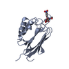
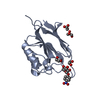
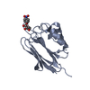
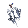
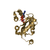
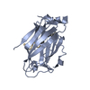
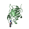
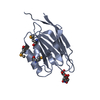
- PDB-5xfc: Serial femtosecond X-ray structure of a stem domain of human O-ma... -
+
Open data
ID or keywords:
Loading...
-
Basic information
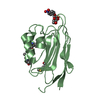
Entry
Database: PDB / ID: 5xfc
Title
Serial femtosecond X-ray structure of a stem domain of human O-mannose beta-1,2-N-acetylglucosaminyltransferase solved by Se-SAD using XFEL (refined against 13,000 patterns)
Components
Protein O-linked-mannose beta-1,2-N-acetylglucosaminyltransferase 1
Keywords
SUGAR BINDING PROTEIN / glycosyltransferase / carbohydrate-binding domain
Function / homology
Function and homology information
beta-1,3-galactosyl-O-glycosyl-glycoprotein beta-1,3-N-acetylglucosaminyltransferase activity / Defective POMGNT1 causes MDDGA3, MDDGB3 and MDDGC3 / DAG1 core M2 glycosylations / DAG1 core M1 glycosylations / Matriglycan biosynthesis on DAG1 / localization of cell / protein O-linked glycosylation via N-acetylgalactosamine / protein O-linked glycosylation via mannose / acetylglucosaminyltransferase activity / reactive gliosis ...beta-1,3-galactosyl-O-glycosyl-glycoprotein beta-1,3-N-acetylglucosaminyltransferase activity / Defective POMGNT1 causes MDDGA3, MDDGB3 and MDDGC3 / DAG1 core M2 glycosylations / DAG1 core M1 glycosylations / Matriglycan biosynthesis on DAG1 / localization of cell / protein O-linked glycosylation via N-acetylgalactosamine / protein O-linked glycosylation via mannose / acetylglucosaminyltransferase activity / reactive gliosis / basement membrane organization / dentate gyrus development / protein O-linked glycosylation / Transferases; Glycosyltransferases; Hexosyltransferases / myelination / sensory perception of sound / manganese ion binding / carbohydrate binding / gene expression / Golgi membrane / membrane Similarity search - Function
In the structure databanks used in Yorodumi, some data are registered as the other names, "COVID-19 virus" and "2019-nCoV". Here are the details of the virus and the list of structure data.
Jan 31, 2019. EMDB accession codes are about to change! (news from PDBe EMDB page)
EMDB accession codes are about to change! (news from PDBe EMDB page)
The allocation of 4 digits for EMDB accession codes will soon come to an end. Whilst these codes will remain in use, new EMDB accession codes will include an additional digit and will expand incrementally as the available range of codes is exhausted. The current 4-digit format prefixed with “EMD-” (i.e. EMD-XXXX) will advance to a 5-digit format (i.e. EMD-XXXXX), and so on. It is currently estimated that the 4-digit codes will be depleted around Spring 2019, at which point the 5-digit format will come into force.
The EM Navigator/Yorodumi systems omit the EMD- prefix.
Related info.:Q: What is EMD? / ID/Accession-code notation in Yorodumi/EM Navigator
Yorodumi is a browser for structure data from EMDB, PDB, SASBDB, etc.
This page is also the successor to EM Navigator detail page, and also detail information page/front-end page for Omokage search.
The word "yorodu" (or yorozu) is an old Japanese word meaning "ten thousand". "mi" (miru) is to see.
Related info.:EMDB / PDB / SASBDB / Comparison of 3 databanks / Yorodumi Search / Aug 31, 2016. New EM Navigator & Yorodumi / Yorodumi Papers / Jmol/JSmol / Function and homology information / Changes in new EM Navigator and Yorodumi
 Movie
Movie Controller

 Yorodumi
 Open data
 Basic information
 Components
 Keywords
 Function and homology information
 Homo sapiens (human)
 X-RAY DIFFRACTION /
  Authors
 Citation
 Structure visualization
 Downloads & links
 Other downloads


 PDBj
PDBj

 Assembly

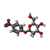
 Type: D-saccharide / Mass: 301.249 Da / Num. of mol.: 2
 Mass: 18.015 Da / Num. of mol.: 171 / Source method: isolated from a natural source / Formula: H2O
 Sample preparation
 / Beamline: BL3 / Wavelength: 0.954 Å
 Processing