 Movie
Movie Controller

[English] 日本語
 Yorodumi
- PDB-5fnx: Crystal structure at pH 9.0 of a potato STI-Kunitz bi-functional ... -
+ Open data

- Basic information
| Entry | Database: PDB / ID: 5fnx | ||||||
|---|---|---|---|---|---|---|---|
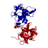
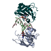
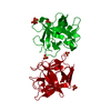
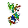
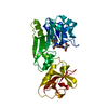
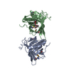
| Title | Crystal structure at pH 9.0 of a potato STI-Kunitz bi-functional inhibitor of serine and aspartic proteases in space group p4322 and ph 9.0 | ||||||
 Components Components | POTATO STI-KUNITZ BI-FUNCTIONAL INHIBITOR E3AD_N19D | ||||||
 Keywords Keywords | HYDROLASE INHIBITOR / HYDROLASE / STI-KUNITZ INHIBITOR / ASPARTIC PROTEASES / SERINE PROTEASES / PROTEASE INHIBITOR / BI-FUNCTIONAL PROTEASE INHIBITOR / KUNITZ-TYPE INHIBITOR | ||||||
| Function / homology |  Function and homology information Function and homology informationaspartic-type endopeptidase inhibitor activity / serine-type endopeptidase inhibitor activity Similarity search - Function | ||||||
| Biological species |  | ||||||
| Method |  X-RAY DIFFRACTION / SYNCHROTRON / MOLECULAR REPLACEMENT / Resolution: 2.65 Å X-RAY DIFFRACTION / SYNCHROTRON / MOLECULAR REPLACEMENT / Resolution: 2.65 Å | ||||||
 Authors Authors | Guerra, Y. / Rudino-Pinera, E. | ||||||
 Citation Citation | Journal: J. Struct. Biol. / Year: 2016 Title: Structures of a bi-functional Kunitz-type STI family inhibitor of serine and aspartic proteases: Could the aspartic protease inhibition have evolved from a canonical serine protease-binding loop? Authors: Guerra, Y. / Valiente, P.A. / Pons, T. / Berry, C. / Rudino-Pinera, E. | ||||||
| History |
|
- Structure visualization
| Structure viewer | Molecule: MolmilJmol/JSmol |
|---|
- Downloads & links
-Download
| PDBx/mmCIF format | 5fnx.cif.gz | 48.4 KB | Display | PDBx/mmCIF format |
|---|---|---|---|---|
| PDB format | pdb5fnx.ent.gz | 34.7 KB | Display | PDB format |
| PDBx/mmJSON format | 5fnx.json.gz | Tree view | PDBx/mmJSON format | |
| Others |  Other downloads Other downloads |
-Validation report
| Arichive directory | https://data.pdbj.org/pub/pdb/validation_reports/fn/5fnxftp://data.pdbj.org/pub/pdb/validation_reports/fn/5fnx | HTTPS FTP |
|---|
-Related structure data
| Related structure data |  5fnwC  5fzuC  5fzyC  5fzzC  5g00C 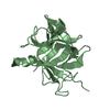 3tc2S C: citing same article ( S: Starting model for refinement |
|---|---|
| Similar structure data |


-Links
 PDBj
PDBj- Assembly
| Deposited unit | 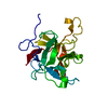
| ||||||||
|---|---|---|---|---|---|---|---|---|---|
| 1 | 
| ||||||||
| Unit cell |
|
-Components
| #1: Protein | Mass: 20527.557 Da / Num. of mol.: 1 Source method: isolated from a genetically manipulated source Details: THE SEQUENCE OF THE POTATO STI-KUNITZ BI- FUNCTIONAL INHIBITOR E3AD_N19D HAS NOT BEEN DEPOSITED AT UNIPROT NOR GENBANK. Source: (gene. exp.)  KOMAGATAELLA PASTORIS GS115 (fungus) / References: UniProt: M1AKE5*PLUS KOMAGATAELLA PASTORIS GS115 (fungus) / References: UniProt: M1AKE5*PLUS |
|---|---|
| #2: Water | ChemComp-HOH /  Mass: 18.015 Da / Num. of mol.: 19 / Source method: isolated from a natural source / Formula: H2O Mass: 18.015 Da / Num. of mol.: 19 / Source method: isolated from a natural source / Formula: H2O |
| Has protein modification | Y |
| Sequence details | WHEN POTATO STI-KUNITZ BI-FUNCTIONAL INHIBITOR E3AD_N19D WAS SEQUENCED RESIDUES 19 AND 177 WERE N ...WHEN POTATO STI-KUNITZ BI-FUNCTIONAL |
-Experimental details
-Experiment
| Experiment | Method: X-RAY DIFFRACTION / Number of used crystals: 1 |
|---|
- Sample preparation
| Crystal | Density Matthews: 3.52 Å3/Da / Density % sol: 65.03 % / Description: NONE |
|---|---|
| Crystal grow | pH: 9 Details: CRYSTAL WAS CRYSTALLIZED FROM 10% MPD, 0.1M BICINE PH 9.0 |
-Data collection
| Diffraction | Mean temperature: 100 K |
|---|---|
| Diffraction source | Source: SYNCHROTRON / Site: APS  / Beamline: 19-ID / Wavelength: 0.9792 / Beamline: 19-ID / Wavelength: 0.9792 |
| Detector | Type: ADSC QUANTUM 315r / Detector: CCD / Date: Oct 24, 2015 / Details: MICROBEAM |
| Radiation | Monochromator: SI(111) / Protocol: SINGLE WAVELENGTH / Monochromatic (M) / Laue (L): M / Scattering type: x-ray |
| Radiation wavelength | Wavelength: 0.9792 Å / Relative weight: 1 |
| Reflection | Resolution: 2.65→19.5 Å / Num. obs: 8941 / % possible obs: 99.4 % / Observed criterion σ(I): 0 / Redundancy: 18.3 % / Biso Wilson estimate: 72.42 Å2 / Rmerge(I) obs: 0.05 / Net I/σ(I): 40.61 |
| Reflection shell | Resolution: 2.65→2.75 Å / Redundancy: 19.24 % / Rmerge(I) obs: 0.57 / Mean I/σ(I) obs: 5.98 / % possible all: 100 |
- Processing
| Software |
| ||||||||||||||||||||||||||||
|---|---|---|---|---|---|---|---|---|---|---|---|---|---|---|---|---|---|---|---|---|---|---|---|---|---|---|---|---|---|
| Refinement | Method to determine structure: MOLECULAR REPLACEMENT Starting model: PDB ENTRY 3TC2 Resolution: 2.65→19.451 Å / SU ML: 0.33 / σ(F): 1.36 / Phase error: 30.18 / Stereochemistry target values: ML
| ||||||||||||||||||||||||||||
| Solvent computation | Shrinkage radii: 0.9 Å / VDW probe radii: 1.11 Å / Solvent model: FLAT BULK SOLVENT MODEL | ||||||||||||||||||||||||||||
| Displacement parameters | Biso mean: 71.5 Å2 | ||||||||||||||||||||||||||||
| Refinement step | Cycle: LAST / Resolution: 2.65→19.451 Å
| ||||||||||||||||||||||||||||
| Refine LS restraints |
| ||||||||||||||||||||||||||||
| LS refinement shell |
|
