 Movie
Movie Controller

[English] 日本語
 Yorodumi
- PDB-3p8k: Crystal Structure of a putative carbon-nitrogen family hydrolase ... -
+ Open data

- Basic information
| Entry | Database: PDB / ID: 3p8k | ||||||
|---|---|---|---|---|---|---|---|
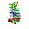
| Title | Crystal Structure of a putative carbon-nitrogen family hydrolase from Staphylococcus aureus | ||||||
 Components Components | Hydrolase, carbon-nitrogen family | ||||||
 Keywords Keywords | HYDROLASE | ||||||
| Function / homology |  Function and homology information Function and homology information | ||||||
| Biological species |  Staphylococcus aureus subsp. aureus COL (bacteria) Staphylococcus aureus subsp. aureus COL (bacteria) | ||||||
| Method |  X-RAY DIFFRACTION / SYNCHROTRON / SAD / Resolution: 1.7 Å X-RAY DIFFRACTION / SYNCHROTRON / SAD / Resolution: 1.7 Å | ||||||
 Authors Authors | Gordon, R.D. / Qiu, W. / Battaile, K. / Lam, K. / Soloveychik, M. / Benetteraj, D. / Romanov, V. / Pai, E.F. / Chirgadze, N.Y. | ||||||
 Citation Citation | Journal: To be Published Title: Crystal Structure of carbon-nitrogen family hydrolase from Staphylococcus aureus Authors: Qiu, W. / Gordon, R.D. / Battaile, K. / Lam, K. / Soloveychik, M. / Benetteraj, D. / Romanov, V. / Pai, E.F. / Chirgadze, N.Y. | ||||||
| History |
|
- Structure visualization
| Structure viewer | Molecule: MolmilJmol/JSmol |
|---|
- Downloads & links
-Download
| PDBx/mmCIF format | 3p8k.cif.gz | 136.3 KB | Display | PDBx/mmCIF format |
|---|---|---|---|---|
| PDB format | pdb3p8k.ent.gz | 105.3 KB | Display | PDB format |
| PDBx/mmJSON format | 3p8k.json.gz | Tree view | PDBx/mmJSON format | |
| Others |  Other downloads Other downloads |
-Validation report
| Summary document | 3p8k_validation.pdf.gz | 480.8 KB | Display | wwPDB validaton report |
|---|---|---|---|---|
| Full document | 3p8k_full_validation.pdf.gz | 486.8 KB | Display | |
| Data in XML | 3p8k_validation.xml.gz | 32.6 KB | Display | |
| Data in CIF | 3p8k_validation.cif.gz | 45.5 KB | Display | |
| Arichive directory | https://data.pdbj.org/pub/pdb/validation_reports/p8/3p8kftp://data.pdbj.org/pub/pdb/validation_reports/p8/3p8k | HTTPS FTP |
-Related structure data
| Similar structure data |
|---|

-Links
 PDBj
PDBj- Assembly
| Deposited unit | 
| ||||||||
|---|---|---|---|---|---|---|---|---|---|
| 1 |
| ||||||||
| Unit cell |
| ||||||||
| Details | THE BIOLOGICAL ASSEMBLY IS UNKNOWN AS PER AUTHORS |
-Components
-Protein , 1 types, 2 molecules AB
| #1: Protein | Mass: 32354.229 Da / Num. of mol.: 2 Source method: isolated from a genetically manipulated source Source: (gene. exp.) Staphylococcus aureus subsp. aureus COL (bacteria)Strain: COL / Gene: SACOL2021 / Production host: |
|---|
-Non-polymers , 6 types, 566 molecules 


| #2: Chemical | ChemComp-TRS /  Mass: 122.143 Da / Num. of mol.: 1 / Source method: obtained synthetically / Formula: C4H12NO3 / Comment: pH buffer*YM Mass: 122.143 Da / Num. of mol.: 1 / Source method: obtained synthetically / Formula: C4H12NO3 / Comment: pH buffer*YM | ||||||||
|---|---|---|---|---|---|---|---|---|---|
| #3: Chemical | ChemComp-CL /  Mass: 35.453 Da / Num. of mol.: 4 / Source method: obtained synthetically / Formula: Cl Mass: 35.453 Da / Num. of mol.: 4 / Source method: obtained synthetically / Formula: Cl#4: Chemical |  Mass: 106.120 Da / Num. of mol.: 3 / Source method: obtained synthetically / Formula: C4H10O3 Mass: 106.120 Da / Num. of mol.: 3 / Source method: obtained synthetically / Formula: C4H10O3#5: Chemical |  Mass: 150.173 Da / Num. of mol.: 2 / Source method: obtained synthetically / Formula: C6H14O4 Mass: 150.173 Da / Num. of mol.: 2 / Source method: obtained synthetically / Formula: C6H14O4#6: Chemical | ChemComp-GOL / |  Mass: 92.094 Da / Num. of mol.: 1 / Source method: obtained synthetically / Formula: C3H8O3 Mass: 92.094 Da / Num. of mol.: 1 / Source method: obtained synthetically / Formula: C3H8O3#7: Water | ChemComp-HOH / | Mass: 18.015 Da / Num. of mol.: 555 / Source method: isolated from a natural source / Formula: H2O |
-Details
| Has protein modification | Y |
|---|
-Experimental details
-Experiment
| Experiment | Method: X-RAY DIFFRACTION / Number of used crystals: 1 |
|---|
- Sample preparation
| Crystal | Density Matthews: 2.18 Å3/Da / Density % sol: 43.49 % |
|---|---|
| Crystal grow | Temperature: 293 K / Method: vapor diffusion / pH: 8.2 Details: 0.2M magnesium chloride, 0.1M TRIS, pH 8.2, 25% PEG 3350, VAPOR DIFFUSION, temperature 293K |
-Data collection
| Diffraction | Mean temperature: 100 K |
|---|---|
| Diffraction source | Source: SYNCHROTRON / Site: APS  / Beamline: 17-ID / Wavelength: 0.9794 Å / Beamline: 17-ID / Wavelength: 0.9794 Å |
| Detector | Type: ADSC QUANTUM 210 / Detector: CCD / Date: Aug 3, 2007 |
| Radiation | Protocol: MAD / Monochromatic (M) / Laue (L): M / Scattering type: x-ray |
| Radiation wavelength | Wavelength: 0.9794 Å / Relative weight: 1 |
| Reflection | Resolution: 1.7→20 Å / Num. all: 62691 / Num. obs: 61246 / % possible obs: 97.9 % / Redundancy: 25 % / Biso Wilson estimate: 20.07 Å2 / Rsym value: 0.0803 / Net I/σ(I): 18.1 |
| Reflection shell | Resolution: 1.7→1.8 Å / Redundancy: 2.6 % / Mean I/σ(I) obs: 2.83 / Num. unique all: 7994 / Rsym value: 0.458 / % possible all: 85.5 |
- Processing
| Software |
| |||||||||||||||||||||||||||||||||||||||||||||||||||||||||||||||||||||||||||||||||||||||||||||||
|---|---|---|---|---|---|---|---|---|---|---|---|---|---|---|---|---|---|---|---|---|---|---|---|---|---|---|---|---|---|---|---|---|---|---|---|---|---|---|---|---|---|---|---|---|---|---|---|---|---|---|---|---|---|---|---|---|---|---|---|---|---|---|---|---|---|---|---|---|---|---|---|---|---|---|---|---|---|---|---|---|---|---|---|---|---|---|---|---|---|---|---|---|---|---|---|---|
| Refinement | Method to determine structure: SAD / Resolution: 1.7→19.96 Å / Cor.coef. Fo:Fc: 0.939 / Cor.coef. Fo:Fc free: 0.9197 / Cross valid method: THROUGHOUT / σ(F): 0
| |||||||||||||||||||||||||||||||||||||||||||||||||||||||||||||||||||||||||||||||||||||||||||||||
| Displacement parameters | Biso mean: 23.55 Å2
| |||||||||||||||||||||||||||||||||||||||||||||||||||||||||||||||||||||||||||||||||||||||||||||||
| Refine analyze | Luzzati coordinate error obs: 0.24 Å | |||||||||||||||||||||||||||||||||||||||||||||||||||||||||||||||||||||||||||||||||||||||||||||||
| Refinement step | Cycle: LAST / Resolution: 1.7→19.96 Å
| |||||||||||||||||||||||||||||||||||||||||||||||||||||||||||||||||||||||||||||||||||||||||||||||
| Refine LS restraints |
| |||||||||||||||||||||||||||||||||||||||||||||||||||||||||||||||||||||||||||||||||||||||||||||||
| LS refinement shell | Resolution: 1.7→1.74 Å / Total num. of bins used: 20
|
