 Movie
Movie Controller

[English] 日本語
 Yorodumi
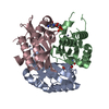
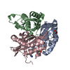
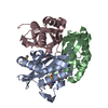
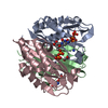
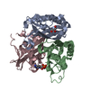
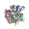
- PDB-3p10: Crystal structure of 2-C-methyl-D-erythritol 2,4-cyclodiphosphate... -
+ Open data

- Basic information
| Entry | Database: PDB / ID: 3p10 | ||||||
|---|---|---|---|---|---|---|---|
| Title | Crystal structure of 2-C-methyl-D-erythritol 2,4-cyclodiphosphate synthase from Burkholderia pseudomallei with cytidine and FOL694, 2-(thiophen-2-yl)phenyl methanol | ||||||
 Components Components | 2-C-methyl-D-erythritol 2,4-cyclodiphosphate synthase | ||||||
 Keywords Keywords | LYASE / Seattle Structural Genomics Center for Infectious Disease / SSGCID / IspF / cytidine / FOL694 / F69 / MEP pathway / isoprene biosynthesis / metal-binding | ||||||
| Function / homology |  Function and homology information Function and homology information2-C-methyl-D-erythritol 2,4-cyclodiphosphate synthase / 2-C-methyl-D-erythritol 2,4-cyclodiphosphate synthase activity / isopentenyl diphosphate biosynthetic process, methylerythritol 4-phosphate pathway / terpenoid biosynthetic process / metal ion binding Similarity search - Function | ||||||
| Biological species |  Burkholderia pseudomallei (bacteria) Burkholderia pseudomallei (bacteria) | ||||||
| Method |  X-RAY DIFFRACTION / MOLECULAR REPLACEMENT / molecular replacement / Resolution: 1.7 Å X-RAY DIFFRACTION / MOLECULAR REPLACEMENT / molecular replacement / Resolution: 1.7 Å | ||||||
 Authors Authors | Seattle Structural Genomics Center for Infectious Disease (SSGCID) | ||||||
 Citation Citation | Journal: J Struct Funct Genomics / Year: 2011 Title: Leveraging structure determination with fragment screening for infectious disease drug targets: MECP synthase from Burkholderia pseudomallei. Authors: Begley, D.W. / Hartley, R.C. / Davies, D.R. / Edwards, T.E. / Leonard, J.T. / Abendroth, J. / Burris, C.A. / Bhandari, J. / Myler, P.J. / Staker, B.L. / Stewart, L.J. #1: Journal: Plos One / Year: 2013Title: Combining functional and structural genomics to sample the essential Burkholderia structome. Authors: Baugh, L. / Gallagher, L.A. / Patrapuvich, R. / Clifton, M.C. / Gardberg, A.S. / Edwards, T.E. / Armour, B. / Begley, D.W. / Dieterich, S.H. / Dranow, D.M. / Abendroth, J. / Fairman, J.W. / ...Authors: Baugh, L. / Gallagher, L.A. / Patrapuvich, R. / Clifton, M.C. / Gardberg, A.S. / Edwards, T.E. / Armour, B. / Begley, D.W. / Dieterich, S.H. / Dranow, D.M. / Abendroth, J. / Fairman, J.W. / Fox, D. / Staker, B.L. / Phan, I. / Gillespie, A. / Choi, R. / Nakazawa-Hewitt, S. / Nguyen, M.T. / Napuli, A. / Barrett, L. / Buchko, G.W. / Stacy, R. / Myler, P.J. / Stewart, L.J. / Manoil, C. / Van Voorhis, W.C. | ||||||
| History |
|
- Structure visualization
| Structure viewer | Molecule: MolmilJmol/JSmol |
|---|
- Downloads & links
-Download
| PDBx/mmCIF format | 3p10.cif.gz | 210.1 KB | Display | PDBx/mmCIF format |
|---|---|---|---|---|
| PDB format | pdb3p10.ent.gz | 167.2 KB | Display | PDB format |
| PDBx/mmJSON format | 3p10.json.gz | Tree view | PDBx/mmJSON format | |
| Others |  Other downloads Other downloads |
-Validation report
| Arichive directory | https://data.pdbj.org/pub/pdb/validation_reports/p1/3p10ftp://data.pdbj.org/pub/pdb/validation_reports/p1/3p10 | HTTPS FTP |
|---|
-Related structure data
| Related structure data | 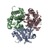 3f0dC 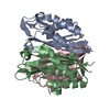 3f0eC  3f0gC 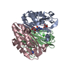 3ieqC  3iewC 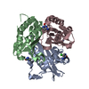 3ikeC 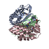 3ikfC 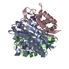 3jvhC 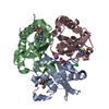 3k14C 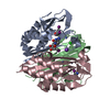 3k2xC 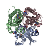 3mbmC 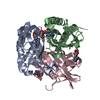 3p0zC 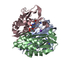 3qhdC C: citing same article ( |
|---|---|
| Similar structure data | |
| Other databases |


-Links
 PDBj
PDBj- Assembly
| Deposited unit | 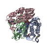
| ||||||||||||
|---|---|---|---|---|---|---|---|---|---|---|---|---|---|
| 1 |
| ||||||||||||
| Unit cell |
| ||||||||||||
| Components on special symmetry positions |
|
-Components
-Protein , 1 types, 3 molecules ABC
| #1: Protein | Mass: 19487.109 Da / Num. of mol.: 3 Source method: isolated from a genetically manipulated source Source: (gene. exp.) Burkholderia pseudomallei (bacteria) / Strain: 1710b / Gene: ispF, BURPS1710b_2511 / Production host: References: UniProt: Q3JRA0, 2-C-methyl-D-erythritol 2,4-cyclodiphosphate synthase |
|---|
-Non-polymers , 6 types, 407 molecules 


| #2: Chemical | ChemComp-CL /  Mass: 35.453 Da / Num. of mol.: 1 / Source method: obtained synthetically / Formula: Cl Mass: 35.453 Da / Num. of mol.: 1 / Source method: obtained synthetically / Formula: Cl | ||||||||
|---|---|---|---|---|---|---|---|---|---|
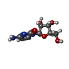
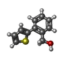
| #3: Chemical |  Mass: 65.409 Da / Num. of mol.: 3 / Source method: obtained synthetically / Formula: Zn Mass: 65.409 Da / Num. of mol.: 3 / Source method: obtained synthetically / Formula: Zn#4: Chemical |  Mass: 243.217 Da / Num. of mol.: 3 / Source method: obtained synthetically / Formula: C9H13N3O5 Mass: 243.217 Da / Num. of mol.: 3 / Source method: obtained synthetically / Formula: C9H13N3O5#5: Chemical | ChemComp-F69 / ( |  Mass: 190.261 Da / Num. of mol.: 1 / Source method: obtained synthetically / Formula: C11H10OS Mass: 190.261 Da / Num. of mol.: 1 / Source method: obtained synthetically / Formula: C11H10OS#6: Chemical | ChemComp-K / |  Mass: 39.098 Da / Num. of mol.: 1 / Source method: obtained synthetically / Formula: K Mass: 39.098 Da / Num. of mol.: 1 / Source method: obtained synthetically / Formula: K#7: Water | ChemComp-HOH / | Mass: 18.015 Da / Num. of mol.: 398 / Source method: isolated from a natural source / Formula: H2O |
-Experimental details
-Experiment
| Experiment | Method: X-RAY DIFFRACTION / Number of used crystals: 1 |
|---|
- Sample preparation
| Crystal | Density Matthews: 2.03 Å3/Da / Density % sol: 39.36 % |
|---|---|
| Crystal grow | Temperature: 289 K / Method: vapor diffusion, sitting drop / pH: 8 Details: 27 mg/mL protein in 20% PEG 4000, 100 mM Tris, 200 mM NaCl, 5 mM ZnCl2. Crystals soaked in 25 mM cytidine and FOL694 in same buffer for 3 weeks, pH 8.0, VAPOR DIFFUSION, SITTING DROP, temperature 289K |
-Data collection
| Diffraction | Mean temperature: 100 K | ||||||||||||||||||||||||||||||||||||||||||||||||||||||||||||||||||||||||||||||||||||||||||||||||||||
|---|---|---|---|---|---|---|---|---|---|---|---|---|---|---|---|---|---|---|---|---|---|---|---|---|---|---|---|---|---|---|---|---|---|---|---|---|---|---|---|---|---|---|---|---|---|---|---|---|---|---|---|---|---|---|---|---|---|---|---|---|---|---|---|---|---|---|---|---|---|---|---|---|---|---|---|---|---|---|---|---|---|---|---|---|---|---|---|---|---|---|---|---|---|---|---|---|---|---|---|---|---|
| Diffraction source | Source: ROTATING ANODE / Type: RIGAKU FR-E+ SUPERBRIGHT / Wavelength: 1.54178 Å | ||||||||||||||||||||||||||||||||||||||||||||||||||||||||||||||||||||||||||||||||||||||||||||||||||||
| Detector | Type: RIGAKU SATURN 944+ / Detector: CCD / Date: Aug 31, 2010 | ||||||||||||||||||||||||||||||||||||||||||||||||||||||||||||||||||||||||||||||||||||||||||||||||||||
| Radiation | Protocol: SINGLE WAVELENGTH / Monochromatic (M) / Laue (L): M / Scattering type: x-ray | ||||||||||||||||||||||||||||||||||||||||||||||||||||||||||||||||||||||||||||||||||||||||||||||||||||
| Radiation wavelength | Wavelength: 1.54178 Å / Relative weight: 1 | ||||||||||||||||||||||||||||||||||||||||||||||||||||||||||||||||||||||||||||||||||||||||||||||||||||
| Reflection | Resolution: 1.7→20 Å / Num. obs: 50700 / % possible obs: 98.4 % / Observed criterion σ(I): -3 / Rmerge(I) obs: 0.022 / Net I/σ(I): 30.42 | ||||||||||||||||||||||||||||||||||||||||||||||||||||||||||||||||||||||||||||||||||||||||||||||||||||
| Reflection shell |
|
-Phasing
| Phasing | Method: molecular replacement |
|---|
- Processing
| Software |
| ||||||||||||||||||||||||||||||||||||||||||||||||||||||||||||||||||||||||||||||||||||||||||||||||||||||||||||||||||||||||||||||||||||||||||||||||||||||||||||||||||||||||||
|---|---|---|---|---|---|---|---|---|---|---|---|---|---|---|---|---|---|---|---|---|---|---|---|---|---|---|---|---|---|---|---|---|---|---|---|---|---|---|---|---|---|---|---|---|---|---|---|---|---|---|---|---|---|---|---|---|---|---|---|---|---|---|---|---|---|---|---|---|---|---|---|---|---|---|---|---|---|---|---|---|---|---|---|---|---|---|---|---|---|---|---|---|---|---|---|---|---|---|---|---|---|---|---|---|---|---|---|---|---|---|---|---|---|---|---|---|---|---|---|---|---|---|---|---|---|---|---|---|---|---|---|---|---|---|---|---|---|---|---|---|---|---|---|---|---|---|---|---|---|---|---|---|---|---|---|---|---|---|---|---|---|---|---|---|---|---|---|---|---|---|---|
| Refinement | Method to determine structure: MOLECULAR REPLACEMENT / Resolution: 1.7→19.91 Å / Cor.coef. Fo:Fc: 0.967 / Cor.coef. Fo:Fc free: 0.953 / Occupancy max: 1 / Occupancy min: 0.3 / SU B: 3.877 / SU ML: 0.058 / SU R Cruickshank DPI: 0.1097 / Cross valid method: THROUGHOUT / ESU R: 0.098 / ESU R Free: 0.096 / Stereochemistry target values: MAXIMUM LIKELIHOOD / Details: HYDROGENS HAVE BEEN ADDED IN THE RIDING POSITIONS
| ||||||||||||||||||||||||||||||||||||||||||||||||||||||||||||||||||||||||||||||||||||||||||||||||||||||||||||||||||||||||||||||||||||||||||||||||||||||||||||||||||||||||||
| Solvent computation | Ion probe radii: 0.8 Å / Shrinkage radii: 0.8 Å / VDW probe radii: 1.4 Å / Solvent model: MASK | ||||||||||||||||||||||||||||||||||||||||||||||||||||||||||||||||||||||||||||||||||||||||||||||||||||||||||||||||||||||||||||||||||||||||||||||||||||||||||||||||||||||||||
| Displacement parameters | Biso mean: 21.331 Å2
| ||||||||||||||||||||||||||||||||||||||||||||||||||||||||||||||||||||||||||||||||||||||||||||||||||||||||||||||||||||||||||||||||||||||||||||||||||||||||||||||||||||||||||
| Refinement step | Cycle: LAST / Resolution: 1.7→19.91 Å
| ||||||||||||||||||||||||||||||||||||||||||||||||||||||||||||||||||||||||||||||||||||||||||||||||||||||||||||||||||||||||||||||||||||||||||||||||||||||||||||||||||||||||||
| Refine LS restraints |
| ||||||||||||||||||||||||||||||||||||||||||||||||||||||||||||||||||||||||||||||||||||||||||||||||||||||||||||||||||||||||||||||||||||||||||||||||||||||||||||||||||||||||||
| LS refinement shell | Resolution: 1.7→1.744 Å / Total num. of bins used: 20
| ||||||||||||||||||||||||||||||||||||||||||||||||||||||||||||||||||||||||||||||||||||||||||||||||||||||||||||||||||||||||||||||||||||||||||||||||||||||||||||||||||||||||||
| Refinement TLS params. | Method: refined / Refine-ID: X-RAY DIFFRACTION
| ||||||||||||||||||||||||||||||||||||||||||||||||||||||||||||||||||||||||||||||||||||||||||||||||||||||||||||||||||||||||||||||||||||||||||||||||||||||||||||||||||||||||||
| Refinement TLS group |
|
