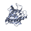
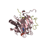
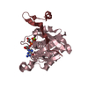
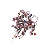
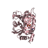
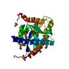
TRANSPORT PROTEIN / Structural Genomics Consortium / SGC / START domain / cholesterol transport / cholesterol / mitochondria
Function / homology
Function and homology information
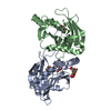
positive regulation of bile acid biosynthetic process / regulation of steroid biosynthetic process / glucocorticoid biosynthetic process / intracellular cholesterol transport / steroid biosynthetic process / Pregnenolone biosynthesis / cholesterol transfer activity / cholesterol binding / cholesterol metabolic process / Mitochondrial protein degradation ...positive regulation of bile acid biosynthetic process / regulation of steroid biosynthetic process / glucocorticoid biosynthetic process / intracellular cholesterol transport / steroid biosynthetic process / Pregnenolone biosynthesis / cholesterol transfer activity / cholesterol binding / cholesterol metabolic process / Mitochondrial protein degradation / mitochondrial intermembrane space / mitochondrial outer membrane / mitochondrial matrix / mitochondrion Similarity search - Function
Steroidogenic acute regulatory protein / Steroidogenic acute regulatory protein-like / in StAR and phosphatidylcholine transfer protein / START domain / START domain / START domain profile. / START domain / Alpha-D-Glucose-1,6-Bisphosphate; Chain A, domain 4 / START-like domain superfamily / 2-Layer Sandwich / Alpha Beta Similarity search - Domain/homology
In the structure databanks used in Yorodumi, some data are registered as the other names, "COVID-19 virus" and "2019-nCoV". Here are the details of the virus and the list of structure data.
Jan 31, 2019. EMDB accession codes are about to change! (news from PDBe EMDB page)
EMDB accession codes are about to change! (news from PDBe EMDB page)
The allocation of 4 digits for EMDB accession codes will soon come to an end. Whilst these codes will remain in use, new EMDB accession codes will include an additional digit and will expand incrementally as the available range of codes is exhausted. The current 4-digit format prefixed with “EMD-” (i.e. EMD-XXXX) will advance to a 5-digit format (i.e. EMD-XXXXX), and so on. It is currently estimated that the 4-digit codes will be depleted around Spring 2019, at which point the 5-digit format will come into force.
The EM Navigator/Yorodumi systems omit the EMD- prefix.
Related info.:Q: What is EMD? / ID/Accession-code notation in Yorodumi/EM Navigator
Yorodumi is a browser for structure data from EMDB, PDB, SASBDB, etc.
This page is also the successor to EM Navigator detail page, and also detail information page/front-end page for Omokage search.
The word "yorodu" (or yorozu) is an old Japanese word meaning "ten thousand". "mi" (miru) is to see.
Related info.:EMDB / PDB / SASBDB / Comparison of 3 databanks / Yorodumi Search / Aug 31, 2016. New EM Navigator & Yorodumi / Yorodumi Papers / Jmol/JSmol / Function and homology information / Changes in new EM Navigator and Yorodumi
 Movie
Movie Controller


 Open data
 Basic information
 Components
 Keywords
 Function and homology information
 Homo sapiens (human)
 X-RAY DIFFRACTION /
  Authors
 Citation
 Structure visualization
 Downloads & links
 Other downloads


 PDBj
PDBj Assembly


 Sample preparation
 / Beamline: ID29 / Wavelength: 0.97948 Å
 Processing