 Movie
Movie Controller


+ Open data

- Basic information
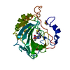
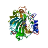
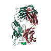
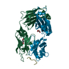
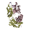
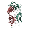
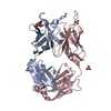
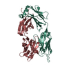
| Entry | Database: PDB / ID: 3okm | ||||||
|---|---|---|---|---|---|---|---|
| Title | Crystal structure of unliganded S25-39 | ||||||
 Components Components |
| ||||||
 Keywords Keywords | IMMUNE SYSTEM / antibody / Fab / IgG / carbohydrate | ||||||
| Function / homology | Immunoglobulins / Immunoglobulin-like / Sandwich / Mainly Beta Function and homology information Function and homology information | ||||||
| Biological species |  | ||||||
| Method |  X-RAY DIFFRACTION / MOLECULAR REPLACEMENT / molecular replacement / Resolution: 2.4 Å X-RAY DIFFRACTION / MOLECULAR REPLACEMENT / molecular replacement / Resolution: 2.4 Å | ||||||
 Authors Authors | Blackler, R.J. / Evans, S.V. | ||||||
 Citation Citation | Journal: Biochemistry / Year: 2011 Title: A Common NH53K Mutation in the Combining Site of Antibodies Raised against Chlamydial LPS Glycoconjugates Significantly Increases Avidity. Authors: Blackler, R.J. / Brooks, C.L. / Evans, D.W. / Brade, L. / Kosma, P. / Brade, H. / Evans, S.V. | ||||||
| History |
|
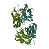
- Structure visualization
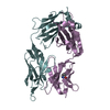
| Structure viewer | Molecule: MolmilJmol/JSmol |
|---|
- Downloads & links
-Download
| PDBx/mmCIF format | 3okm.cif.gz | 100 KB | Display | PDBx/mmCIF format |
|---|---|---|---|---|
| PDB format | pdb3okm.ent.gz | 74.7 KB | Display | PDB format |
| PDBx/mmJSON format | 3okm.json.gz | Tree view | PDBx/mmJSON format | |
| Others |  Other downloads Other downloads |
-Validation report
| Summary document | 3okm_validation.pdf.gz | 436.7 KB | Display | wwPDB validaton report |
|---|---|---|---|---|
| Full document | 3okm_full_validation.pdf.gz | 446 KB | Display | |
| Data in XML | 3okm_validation.xml.gz | 22.3 KB | Display | |
| Data in CIF | 3okm_validation.cif.gz | 29.6 KB | Display | |
| Arichive directory | https://data.pdbj.org/pub/pdb/validation_reports/ok/3okmftp://data.pdbj.org/pub/pdb/validation_reports/ok/3okm | HTTPS FTP |
-Related structure data
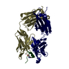
| Related structure data | 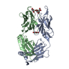 3okdC 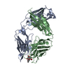 3okeC 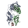 3okkC 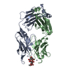 3oklC 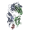 3oknC 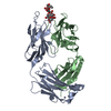 3okoC C: citing same article ( |
|---|---|
| Similar structure data |


-Links
 PDBj
PDBj

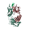
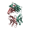
- Assembly
| Deposited unit | 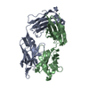
| ||||||||
|---|---|---|---|---|---|---|---|---|---|
| 1 |
| ||||||||
| Unit cell |
|
-Components
| #1: Antibody | Mass: 24182.846 Da / Num. of mol.: 1 / Source method: isolated from a natural source / Details: ascites / Source: (natural) |
|---|---|
| #2: Antibody | Mass: 24022.961 Da / Num. of mol.: 1 / Source method: isolated from a natural source / Details: ascites / Source: (natural) |
| #3: Water | ChemComp-HOH /  Mass: 18.015 Da / Num. of mol.: 175 / Source method: isolated from a natural source / Formula: H2O Mass: 18.015 Da / Num. of mol.: 175 / Source method: isolated from a natural source / Formula: H2O |
| Has protein modification | Y |
-Experimental details
-Experiment
| Experiment | Method: X-RAY DIFFRACTION / Number of used crystals: 1 |
|---|
- Sample preparation
| Crystal | Density Matthews: 2.44 Å3/Da / Density % sol: 49.55 % |
|---|---|
| Crystal grow | Temperature: 289 K / Method: hanging drop / pH: 6.5 Details: PEG 3350, MPD, ZnAc, NaCacod, pH 6.5, hanging drop, temperature 289K |
-Data collection
| Diffraction | Mean temperature: 100 K | |||||||||||||||||||||||||||||||||||||||||||||||||||||||
|---|---|---|---|---|---|---|---|---|---|---|---|---|---|---|---|---|---|---|---|---|---|---|---|---|---|---|---|---|---|---|---|---|---|---|---|---|---|---|---|---|---|---|---|---|---|---|---|---|---|---|---|---|---|---|---|---|
| Diffraction source | Source: ROTATING ANODE / Type: RIGAKU MICROMAX-002 / Wavelength: 1.5418 Å | |||||||||||||||||||||||||||||||||||||||||||||||||||||||
| Detector | Type: RIGAKU RAXIS / Detector: IMAGE PLATE / Date: Jun 8, 2010 / Details: OSMIC BLUE MIRRORS | |||||||||||||||||||||||||||||||||||||||||||||||||||||||
| Radiation | Protocol: SINGLE WAVELENGTH / Monochromatic (M) / Laue (L): M / Scattering type: x-ray | |||||||||||||||||||||||||||||||||||||||||||||||||||||||
| Radiation wavelength | Wavelength: 1.5418 Å / Relative weight: 1 | |||||||||||||||||||||||||||||||||||||||||||||||||||||||
| Reflection | Resolution: 2.4→19.91 Å / Num. obs: 16790 / % possible obs: 92.6 % / Redundancy: 2.95 % / Rmerge(I) obs: 0.097 / Net I/σ(I): 7.4 | |||||||||||||||||||||||||||||||||||||||||||||||||||||||
| Reflection shell |
|
-Phasing
| Phasing | Method: molecular replacement | |||||||||
|---|---|---|---|---|---|---|---|---|---|---|
| Phasing MR | Rfactor: 44.74 / Model details: Phaser MODE: MR_AUTO
|
- Processing
| Software |
| |||||||||||||||||||||||||||||||||||||||||||||||||
|---|---|---|---|---|---|---|---|---|---|---|---|---|---|---|---|---|---|---|---|---|---|---|---|---|---|---|---|---|---|---|---|---|---|---|---|---|---|---|---|---|---|---|---|---|---|---|---|---|---|---|
| Refinement | Method to determine structure: MOLECULAR REPLACEMENT / Resolution: 2.4→19.766 Å / Occupancy max: 1 / Occupancy min: 0.41 / SU ML: 0.42 / σ(F): 1.48 / Phase error: 29.53 / Stereochemistry target values: ML
| |||||||||||||||||||||||||||||||||||||||||||||||||
| Solvent computation | Shrinkage radii: 0.83 Å / VDW probe radii: 1.1 Å / Solvent model: FLAT BULK SOLVENT MODEL / Bsol: 62.431 Å2 / ksol: 0.317 e/Å3 | |||||||||||||||||||||||||||||||||||||||||||||||||
| Displacement parameters |
| |||||||||||||||||||||||||||||||||||||||||||||||||
| Refinement step | Cycle: LAST / Resolution: 2.4→19.766 Å
| |||||||||||||||||||||||||||||||||||||||||||||||||
| Refine LS restraints |
| |||||||||||||||||||||||||||||||||||||||||||||||||
| LS refinement shell |
|
