[English] 日本語
Yorodumi
- PDB-3nfw: Crystal structure of nitrilotriacetate monooxygenase component B ... -

+
Open data


ID or keywords:

Loading...

-
Basic information

Entry
Database: PDB / ID: 3nfw
TitleCrystal structure of nitrilotriacetate monooxygenase component B (A0R521 homolog) from Mycobacterium thermoresistibile
ComponentsFlavin reductase-like, FMN-binding protein
KeywordsOXIDOREDUCTASE / Seattle Structural Genomics Center for Infectious Disease / SSGCID / Mycobacterium / tuberculosis / thermoresistible / biodegredation / NADH dependent
Function / homology
Function and homology information


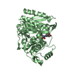
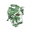
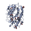
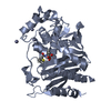
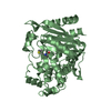
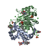
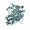
Oxidoreductases; Acting on paired donors, with incorporation or reduction of molecular oxygen; With NADH or NADPH as one donor, and incorporation of one atom of oxygen into the other donor / monooxygenase activity / FMN binding
Similarity search - Function
Flavin reductase like domain / Flavin reductase like domain / Flavin reductase like domain / Electron Transport, Fmn-binding Protein; Chain A / Pnp Oxidase; Chain A / FMN-binding split barrel / Roll / Mainly Beta
Similarity search - Domain/homology
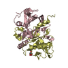
Nitrilotriacetate monooxygenase component B / Flavin reductase-like, FMN-binding protein
Similarity search - Component
Biological speciesMycobacterium thermoresistibile (bacteria)
MethodX-RAY DIFFRACTION / SYNCHROTRON / MOLECULAR REPLACEMENT / molecular replacement / Resolution: 1.6 Å
AuthorsSeattle Structural Genomics Center for Infectious Disease (SSGCID)
Citation
Journal: Acta Crystallogr.,Sect.F / Year: 2011
Title: Structure of nitrilotriacetate monooxygenase component B from Mycobacterium thermoresistibile.
Authors: Zhang, Y. / Edwards, T.E. / Begley, D.W. / Abramov, A. / Thompkins, K.B. / Ferrell, M. / Guo, W.J. / Phan, I. / Olsen, C. / Napuli, A. / Sankaran, B. / Stacy, R. / Van Voorhis, W.C. / Stewart, L.J. / Myler, P.J.
#1: Journal: Tuberculosis (Edinb) / Year: 2015
Title: Increasing the structural coverage of tuberculosis drug targets.
Authors: Baugh, L. / Phan, I. / Begley, D.W. / Clifton, M.C. / Armour, B. / Dranow, D.M. / Taylor, B.M. / Muruthi, M.M. / Abendroth, J. / Fairman, J.W. / Fox, D. / Dieterich, S.H. / Staker, B.L. / ...Authors: Baugh, L. / Phan, I. / Begley, D.W. / Clifton, M.C. / Armour, B. / Dranow, D.M. / Taylor, B.M. / Muruthi, M.M. / Abendroth, J. / Fairman, J.W. / Fox, D. / Dieterich, S.H. / Staker, B.L. / Gardberg, A.S. / Choi, R. / Hewitt, S.N. / Napuli, A.J. / Myers, J. / Barrett, L.K. / Zhang, Y. / Ferrell, M. / Mundt, E. / Thompkins, K. / Tran, N. / Lyons-Abbott, S. / Abramov, A. / Sekar, A. / Serbzhinskiy, D. / Lorimer, D. / Buchko, G.W. / Stacy, R. / Stewart, L.J. / Edwards, T.E. / Van Voorhis, W.C. / Myler, P.J.
History
DepositionJun 10, 2010Deposition site: RCSB / Processing site: RCSB
Revision 1.0Jun 23, 2010Provider: repository / Type: Initial release
Revision 1.1Jul 13, 2011Group: Version format compliance
Revision 1.2Sep 21, 2011Group: Database references
Revision 1.3Dec 19, 2012Group: Database references / Source and taxonomy / Structure summary
Revision 1.4Apr 22, 2015Group: Database references
Revision 1.5Sep 6, 2023Group: Data collection / Database references ...Data collection / Database references / Derived calculations / Refinement description
Category: chem_comp_atom / chem_comp_bond ...chem_comp_atom / chem_comp_bond / database_2 / pdbx_initial_refinement_model / struct_ref_seq_dif / struct_site
Item: _database_2.pdbx_DOI / _database_2.pdbx_database_accession ..._database_2.pdbx_DOI / _database_2.pdbx_database_accession / _struct_ref_seq_dif.details / _struct_site.pdbx_auth_asym_id / _struct_site.pdbx_auth_comp_id / _struct_site.pdbx_auth_seq_id

-
Structure visualization

Structure viewerMolecule:
MolmilJmol/JSmol

Downloads & links

-
Assembly

Deposited unit
A: Flavin reductase-like, FMN-binding protein
B: Flavin reductase-like, FMN-binding protein
C: Flavin reductase-like, FMN-binding protein
D: Flavin reductase-like, FMN-binding protein
hetero molecules


Theoretical massNumber of molelcules
Total (without water)93,3215
Polymers93,2294
Non-polymers921
Water9,872548
1
A: Flavin reductase-like, FMN-binding protein
B: Flavin reductase-like, FMN-binding protein


Theoretical massNumber of molelcules
Total (without water)46,6152
Polymers46,6152
Non-polymers00
Water362
TypeNameSymmetry operationNumber
identity operation1_555x,y,z1
Buried area8610 Å2
ΔGint-56 kcal/mol
Surface area15290 Å2
MethodPISA
2
C: Flavin reductase-like, FMN-binding protein
D: Flavin reductase-like, FMN-binding protein
hetero molecules


Theoretical massNumber of molelcules
Total (without water)46,7073
Polymers46,6152
Non-polymers921
Water362
TypeNameSymmetry operationNumber
identity operation1_555x,y,z1
Buried area8760 Å2
ΔGint-55 kcal/mol
Surface area15270 Å2
MethodPISA
Unit cell
Length a, b, c (Å)48.450, 91.020, 165.640
Angle α, β, γ (deg.)90.00, 90.00, 90.00
Int Tables number19
Space group name H-MP212121

-
Components

#1: Protein
Flavin reductase-like, FMN-binding protein / nitrilotriacetate monooxygenase component B


Mass: 23307.271 Da / Num. of mol.: 4
Source method: isolated from a genetically manipulated source
Source: (gene. exp.) Mycobacterium thermoresistibile (bacteria)
Production host: Escherichia coli (E. coli) / References: UniProt: G7CEX2, UniProt: E5Q9D7*PLUS
#2: Chemical ChemComp-GOL / GLYCEROL / GLYCERIN / PROPANE-1,2,3-TRIOL


Mass: 92.094 Da / Num. of mol.: 1 / Source method: obtained synthetically / Formula: C3H8O3
#3: Water ChemComp-HOH / water


Mass: 18.015 Da / Num. of mol.: 548 / Source method: isolated from a natural source / Formula: H2O

-
Experimental details

-
Experiment

ExperimentMethod: X-RAY DIFFRACTION / Number of used crystals: 1

-
Sample preparation

CrystalDensity Matthews: 1.96 Å3/Da / Density % sol: 37.21 %
Crystal growTemperature: 289 K / Method: vapor diffusion, sitting drop / pH: 6
Details: 68.9 mg/mL protein against PACT screen condition B10, 0.2 M MgCl2, 0.1 M MES pH 6.0, 20% PEG 6000 with 20% glycerol as cryo-protectant, crystal tracking ID 215735b10, VAPOR DIFFUSION, ...Details: 68.9 mg/mL protein against PACT screen condition B10, 0.2 M MgCl2, 0.1 M MES pH 6.0, 20% PEG 6000 with 20% glycerol as cryo-protectant, crystal tracking ID 215735b10, VAPOR DIFFUSION, SITTING DROP, temperature 289K

-
Data collection

DiffractionMean temperature: 100 K
Diffraction sourceSource: SYNCHROTRON / Site: ALS / Beamline: 5.0.2 / Wavelength: 1 Å
DetectorType: ADSC QUANTUM 315r / Detector: CCD / Date: May 29, 2010
RadiationProtocol: SINGLE WAVELENGTH / Monochromatic (M) / Laue (L): M / Scattering type: x-ray
Radiation wavelengthWavelength: 1 Å / Relative weight: 1
ReflectionResolution: 1.6→50 Å / Num. all: 97543 / Num. obs: 97350 / % possible obs: 99.8 % / Observed criterion σ(I): -3 / Redundancy: 14.5 % / Biso Wilson estimate: 20.998 Å2 / Rmerge(I) obs: 0.095 / Net I/σ(I): 14.4
Reflection shell

Diffraction-ID: 1

Resolution (Å)Highest resolution (Å)Redundancy (%)Rmerge(I) obsMean I/σ(I) obsNum. measured obsNum. unique obs% possible all
1.6-1.6413.70.4285.455343710299.9
1.64-1.690.3656.155237695899.9
1.69-1.740.3057.2548916756100
1.74-1.790.278546026581100
1.79-1.850.2289.2538486361100
1.85-1.910.19810.5526176161100
1.91-1.980.16412.4515745987100
1.98-2.070.13614.349253571599.9
2.07-2.160.12215.847363551099.9
2.16-2.260.10817.545030527999.9
2.26-2.390.10518.242482504599.9
2.39-2.530.09618.740124477999.9
2.53-2.70.0919.636884450699.8
2.7-2.920.08220.833530415099.8
2.92-3.20.07722.230947390099.8
3.2-3.580.07124.226867351399.9
3.58-4.130.06725.323718313199.6
4.13-5.060.06126.721064266299.4
5.06-7.160.05825.416670211199.8
7.160.04825.28470114391.9

-
Phasing

PhasingMethod: molecular replacement
Phasing MRRfactor: 61.42 / Model details: Phaser MODE: MR_AUTO
Highest resolutionLowest resolution
Rotation2.5 Å19.55 Å
Translation2.5 Å19.55 Å

-
Processing

Software
NameVersionClassificationNB
XSCALEdata scaling
PHASER2.1.4phasing
REFMACrefinement
PDB_EXTRACT3.1data extraction
XDSdata reduction
RefinementMethod to determine structure: MOLECULAR REPLACEMENT
Starting model: PDB entry 1rz0
Resolution: 1.6→19.56 Å / Cor.coef. Fo:Fc: 0.955 / Cor.coef. Fo:Fc free: 0.94 / WRfactor Rfree: 0.2239 / WRfactor Rwork: 0.1915 / Occupancy max: 1 / Occupancy min: 0.3 / FOM work R set: 0.8626 / SU B: 3.142 / SU ML: 0.052 / SU R Cruickshank DPI: 0.0913 / SU Rfree: 0.0913 / Cross valid method: THROUGHOUT / σ(F): 0 / ESU R Free: 0.091 / Stereochemistry target values: MAXIMUM LIKELIHOOD
Details: HYDROGENS HAVE BEEN ADDED IN THE RIDING POSITIONS U VALUES: WITH TLS ADDED
RfactorNum. reflection% reflectionSelection details
Rfree0.2175 4857 5 %RANDOM
Rwork0.1864 ---
obs0.188 97026 99.48 %-
Solvent computationIon probe radii: 0.8 Å / Shrinkage radii: 0.8 Å / VDW probe radii: 1.4 Å / Solvent model: MASK
Displacement parametersBiso max: 58.9 Å2 / Biso mean: 16.7061 Å2 / Biso min: 5.32 Å2
Baniso -1Baniso -2Baniso -3
1--0.56 Å20 Å20 Å2
2--0.19 Å20 Å2
3---0.37 Å2
Refinement stepCycle: LAST / Resolution: 1.6→19.56 Å
ProteinNucleic acidLigandSolventTotal
Num. atoms5541 0 6 548 6095
Refine LS restraints
Refine-IDTypeDev idealDev ideal targetNumber
X-RAY DIFFRACTIONr_bond_refined_d0.0150.0225719
X-RAY DIFFRACTIONr_angle_refined_deg1.2731.9387796
X-RAY DIFFRACTIONr_dihedral_angle_1_deg6.5575728
X-RAY DIFFRACTIONr_dihedral_angle_2_deg33.40822.68250
X-RAY DIFFRACTIONr_dihedral_angle_3_deg11.0115843
X-RAY DIFFRACTIONr_dihedral_angle_4_deg17.4731542
X-RAY DIFFRACTIONr_chiral_restr0.0960.2864
X-RAY DIFFRACTIONr_gen_planes_refined0.0060.0214440
X-RAY DIFFRACTIONr_mcbond_it0.7381.53616
X-RAY DIFFRACTIONr_mcangle_it1.34625830
X-RAY DIFFRACTIONr_scbond_it2.01732103
X-RAY DIFFRACTIONr_scangle_it3.334.51962
LS refinement shellResolution: 1.6→1.642 Å / Total num. of bins used: 20
RfactorNum. reflection% reflection
Rfree0.227 375 -
Rwork0.18 6691 -
all-7066 -
obs--99.58 %
Refinement TLS params.

Method: refined / Refine-ID: X-RAY DIFFRACTION

IDL112)L122)L132)L222)L232)L332)S11 (Å °)S12 (Å °)S13 (Å °)S21 (Å °)S22 (Å °)S23 (Å °)S31 (Å °)S32 (Å °)S33 (Å °)T112)T122)T132)T222)T232)T332)Origin x (Å)Origin y (Å)Origin z (Å)
10.11130.08160.00880.1616-0.03560.22020.0027-0.0047-0.01990.0157-0.0068-0.00440.0016-0.01720.00410.0081-0.0028-0.00670.00810.00550.01817.08597.8325.7255
20.18770.11-0.02890.17750.01170.1694-0.00330.0122-0.0137-0.0116-0.0003-0.0159-0.0313-0.00160.00360.00720.0001-0.00080.0210.00620.005325.430817.521113.8698
30.17950.0497-0.04120.151-0.01350.2362-0.0068-0.0037-0.02810.0237-0.00380.00420.0073-0.00960.01060.0109-0.0033-0.00380.00570.00610.016413.0925-37.654525.9875
40.23610.07920.01110.1166-0.02840.1805-0.00550.0219-0.0037-0.00680.0041-0.0149-0.01490.00450.00140.0067-0.0007-0.00150.02150.00750.006421.2226-28.179514.1649
Refinement TLS group
IDRefine-IDRefine TLS-IDAuth asym-IDAuth seq-ID
1X-RAY DIFFRACTION1A3 - 181
2X-RAY DIFFRACTION2B1 - 181
3X-RAY DIFFRACTION3C2 - 181
4X-RAY DIFFRACTION4D1 - 182

+
About Yorodumi

-
News

-
Feb 9, 2022. New format data for meta-information of EMDB entries

New format data for meta-information of EMDB entries

  • Version 3 of the EMDB header file is now the official format.
  • The previous official version 1.9 will be removed from the archive.

Related info.:EMDB header

External links:wwPDB to switch to version 3 of the EMDB data model

-
Aug 12, 2020. Covid-19 info

Covid-19 info

URL: https://pdbjlvh1.pdbj.org/emnavi/covid19.php

New page: Covid-19 featured information page in EM Navigator.

Related info.:Covid-19 info / Mar 5, 2020. Novel coronavirus structure data

+
Mar 5, 2020. Novel coronavirus structure data

Novel coronavirus structure data

Related info.:Yorodumi Speices / Aug 12, 2020. Covid-19 info

External links:COVID-19 featured content - PDBj / Molecule of the Month (242):Coronavirus Proteases

+
Jan 31, 2019. EMDB accession codes are about to change! (news from PDBe EMDB page)

EMDB accession codes are about to change! (news from PDBe EMDB page)

  • The allocation of 4 digits for EMDB accession codes will soon come to an end. Whilst these codes will remain in use, new EMDB accession codes will include an additional digit and will expand incrementally as the available range of codes is exhausted. The current 4-digit format prefixed with “EMD-” (i.e. EMD-XXXX) will advance to a 5-digit format (i.e. EMD-XXXXX), and so on. It is currently estimated that the 4-digit codes will be depleted around Spring 2019, at which point the 5-digit format will come into force.
  • The EM Navigator/Yorodumi systems omit the EMD- prefix.

Related info.:Q: What is EMD? / ID/Accession-code notation in Yorodumi/EM Navigator

External links:EMDB Accession Codes are Changing Soon! / Contact to PDBj

+
Jul 12, 2017. Major update of PDB

Major update of PDB

  • wwPDB released updated PDB data conforming to the new PDBx/mmCIF dictionary.
  • This is a major update changing the version number from 4 to 5, and with Remediation, in which all the entries are updated.
  • In this update, many items about electron microscopy experimental information are reorganized (e.g. em_software).
  • Now, EM Navigator and Yorodumi are based on the updated data.

External links:wwPDB Remediation / Enriched Model Files Conforming to OneDep Data Standards Now Available in the PDB FTP Archive

-
Yorodumi

Thousand views of thousand structures

  • Yorodumi is a browser for structure data from EMDB, PDB, SASBDB, etc.
  • This page is also the successor to EM Navigator detail page, and also detail information page/front-end page for Omokage search.
  • The word "yorodu" (or yorozu) is an old Japanese word meaning "ten thousand". "mi" (miru) is to see.

Related info.:EMDB / PDB / SASBDB / Comparison of 3 databanks / Yorodumi Search / Aug 31, 2016. New EM Navigator & Yorodumi / Yorodumi Papers / Jmol/JSmol / Function and homology information / Changes in new EM Navigator and Yorodumi

Read more