- PDB-6bnc: Crystal structure of the intrinsic colistin resistance enzyme ICR... -
+
Open data
ID or keywords:
Loading...
-
Basic information
Entry
Database: PDB / ID: 6bnc
Title
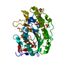
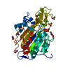
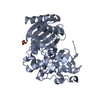
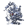
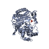
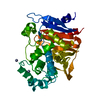
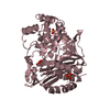
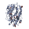
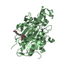
Crystal structure of the intrinsic colistin resistance enzyme ICR(Mc) from Moraxella catarrhalis, catalytic domain, Thr315Ala mutant di-zinc and PEG complex
Components
Phosphoethanolamine transferase
Keywords
TRANSFERASE / antibiotic resistance / colistin / polymycin / phosphoethanolamine transferase / sulfatase fold / alpha/beta protein / Structural Genomics / Center for Structural Genomics of Infectious Diseases / CSGID
Function / homology
Function and homology information
phosphotransferase activity, phosphate group as acceptor / lipopolysaccharide core region biosynthetic process / metal ion binding / plasma membrane Similarity search - Function
Mass: 18.015 Da / Num. of mol.: 1043 / Source method: isolated from a natural source / Formula: H2O
Has protein modification
Y
-
Experimental details
-
Experiment
Experiment
Method: X-RAY DIFFRACTION / Number of used crystals: 1
-
Sample preparation
Crystal
Density Matthews: 2.5 Å3/Da / Density % sol: 50.77 %
Crystal grow
Temperature: 298 K / Method: vapor diffusion, sitting drop / pH: 8 Details: 0.1 M Tris pH 8, 28% (w/v) PEG 4k, 3 mM zinc chloride and 2 mM lipid A headgroup (Glycobiotech GmbH). Cryoprotectant paratone.
In the structure databanks used in Yorodumi, some data are registered as the other names, "COVID-19 virus" and "2019-nCoV". Here are the details of the virus and the list of structure data.
Jan 31, 2019. EMDB accession codes are about to change! (news from PDBe EMDB page)
EMDB accession codes are about to change! (news from PDBe EMDB page)
The allocation of 4 digits for EMDB accession codes will soon come to an end. Whilst these codes will remain in use, new EMDB accession codes will include an additional digit and will expand incrementally as the available range of codes is exhausted. The current 4-digit format prefixed with “EMD-” (i.e. EMD-XXXX) will advance to a 5-digit format (i.e. EMD-XXXXX), and so on. It is currently estimated that the 4-digit codes will be depleted around Spring 2019, at which point the 5-digit format will come into force.
The EM Navigator/Yorodumi systems omit the EMD- prefix.
Related info.:Q: What is EMD? / ID/Accession-code notation in Yorodumi/EM Navigator
Yorodumi is a browser for structure data from EMDB, PDB, SASBDB, etc.
This page is also the successor to EM Navigator detail page, and also detail information page/front-end page for Omokage search.
The word "yorodu" (or yorozu) is an old Japanese word meaning "ten thousand". "mi" (miru) is to see.
Related info.:EMDB / PDB / SASBDB / Comparison of 3 databanks / Yorodumi Search / Aug 31, 2016. New EM Navigator & Yorodumi / Yorodumi Papers / Jmol/JSmol / Function and homology information / Changes in new EM Navigator and Yorodumi
 Movie
Movie Controller

 Yorodumi
 Open data
 Basic information
 Components
 Keywords
 Function and homology information
 Moraxella sp. HMSC061H09 (bacteria)
 X-RAY DIFFRACTION /
  Authors
 United States, 2items
  Citation
 Structure visualization
 Downloads & links
 Other downloads
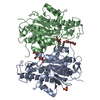

 PDBj
PDBj
 Assembly
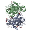

 Mass: 65.409 Da / Num. of mol.: 4 / Source method: obtained synthetically / Formula: Zn / Feature type: SUBJECT OF INVESTIGATION
 Mass: 1529.829 Da / Num. of mol.: 9 / Source method: obtained synthetically / Formula: C69H140O35 / Comment: precipitant*YM
 Mass: 35.453 Da / Num. of mol.: 1 / Source method: obtained synthetically / Formula: Cl
 Mass: 18.015 Da / Num. of mol.: 1043 / Source method: isolated from a natural source / Formula: H2O
 Sample preparation
 Processing