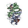
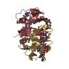
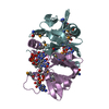
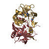
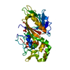
| 登録構造単位 | A: phenol 2-hydroxylase component B
B: phenol 2-hydroxylase component B
C: phenol 2-hydroxylase component B
D: phenol 2-hydroxylase component B
E: phenol 2-hydroxylase component B
F: phenol 2-hydroxylase component B
G: phenol 2-hydroxylase component B
H: phenol 2-hydroxylase component B
ヘテロ分子
| 分子量 (理論値) | 分子数 |
|---|
| 合計 (水以外) | 149,946 | 16 |
|---|
| ポリマ- | 143,661 | 8 |
|---|
| 非ポリマー | 6,284 | 8 |
|---|
| 水 | 7,332 | 407 |
|---|
|
|---|
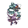
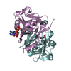
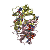
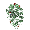
| 1 | A: phenol 2-hydroxylase component B
B: phenol 2-hydroxylase component B
ヘテロ分子
| 分子量 (理論値) | 分子数 |
|---|
| 合計 (水以外) | 37,486 | 4 |
|---|
| ポリマ- | 35,915 | 2 |
|---|
| 非ポリマー | 1,571 | 2 |
|---|
| 水 | 36 | 2 |
|---|
| タイプ | 名称 | 対称操作 | 数 |
|---|
| identity operation | 1_555 | x,y,z | 1 |
| Buried area | 7130 Å2 |
|---|
| ΔGint | -60 kcal/mol |
|---|
| Surface area | 13250 Å2 |
|---|
| 手法 | PISA |
|---|
|
|---|
| 2 | C: phenol 2-hydroxylase component B
D: phenol 2-hydroxylase component B
ヘテロ分子
| 分子量 (理論値) | 分子数 |
|---|
| 合計 (水以外) | 37,486 | 4 |
|---|
| ポリマ- | 35,915 | 2 |
|---|
| 非ポリマー | 1,571 | 2 |
|---|
| 水 | 36 | 2 |
|---|
| タイプ | 名称 | 対称操作 | 数 |
|---|
| identity operation | 1_555 | x,y,z | 1 |
| Buried area | 7170 Å2 |
|---|
| ΔGint | -61 kcal/mol |
|---|
| Surface area | 13220 Å2 |
|---|
| 手法 | PISA |
|---|
|
|---|
| 3 | E: phenol 2-hydroxylase component B
F: phenol 2-hydroxylase component B
ヘテロ分子
| 分子量 (理論値) | 分子数 |
|---|
| 合計 (水以外) | 37,486 | 4 |
|---|
| ポリマ- | 35,915 | 2 |
|---|
| 非ポリマー | 1,571 | 2 |
|---|
| 水 | 36 | 2 |
|---|
| タイプ | 名称 | 対称操作 | 数 |
|---|
| identity operation | 1_555 | x,y,z | 1 |
| Buried area | 7160 Å2 |
|---|
| ΔGint | -61 kcal/mol |
|---|
| Surface area | 13260 Å2 |
|---|
| 手法 | PISA |
|---|
|
|---|
| 4 | G: phenol 2-hydroxylase component B
H: phenol 2-hydroxylase component B
ヘテロ分子
| 分子量 (理論値) | 分子数 |
|---|
| 合計 (水以外) | 37,486 | 4 |
|---|
| ポリマ- | 35,915 | 2 |
|---|
| 非ポリマー | 1,571 | 2 |
|---|
| 水 | 36 | 2 |
|---|
| タイプ | 名称 | 対称操作 | 数 |
|---|
| identity operation | 1_555 | x,y,z | 1 |
| Buried area | 7140 Å2 |
|---|
| ΔGint | -60 kcal/mol |
|---|
| Surface area | 13250 Å2 |
|---|
| 手法 | PISA |
|---|
|
|---|
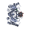
| 単位格子 | | Length a, b, c (Å) | 53.544, 154.268, 83.866 |
|---|
| Angle α, β, γ (deg.) | 90.00, 91.27, 90.00 |
|---|
| Int Tables number | 4 |
|---|
| Space group name H-M | P1211 |
|---|
|
|---|
| 非結晶学的対称性 (NCS) | NCSドメイン: | ID | Ens-ID | 詳細 (eV) |
|---|
| 1 | 1 | A| 2 | 1 | B| 3 | 1 | C| 4 | 1 | D| 5 | 1 | E| 6 | 1 | F| 7 | 1 | G| 8 | 1 | H| 9 | 1 | A| 10 | 1 | B| 11 | 1 | C| 12 | 1 | D| 13 | 1 | E| 14 | 1 | F| 15 | 1 | G| 16 | 1 | H| 17 | 1 | A| 18 | 1 | B| 19 | 1 | C| 20 | 1 | D| 21 | 1 | E| 22 | 1 | F| 23 | 1 | G| 24 | 1 | H | | | | | | | | | | | | | | | | | | | | | | | |
NCSドメイン領域: Ens-ID: 1 / Refine code: 1 | Dom-ID | Component-ID | Beg auth comp-ID | Beg label comp-ID | End auth comp-ID | End label comp-ID | Auth asym-ID | Label asym-ID | Auth seq-ID | Label seq-ID |
|---|
| 1 | 1 | ASPASPGLYGLYAA| 3 - 84 | 3 - 84 | | 2 | 1 | ASPASPGLYGLYBB| 3 - 84 | 3 - 84 | | 3 | 1 | ASPASPGLYGLYCC| 3 - 84 | 3 - 84 | | 4 | 1 | ASPASPGLYGLYDD| 3 - 84 | 3 - 84 | | 5 | 1 | ASPASPGLYGLYEE| 3 - 84 | 3 - 84 | | 6 | 1 | ASPASPGLYGLYFF| 3 - 84 | 3 - 84 | | 7 | 1 | ASPASPGLYGLYGG| 3 - 84 | 3 - 84 | | 8 | 1 | ASPASPGLYGLYHH| 3 - 84 | 3 - 84 | | 9 | 2 | ASPASPGLNGLNAA| 91 - 153 | 91 - 153 | | 10 | 2 | ASPASPGLNGLNBB| 91 - 153 | 91 - 153 | | 11 | 2 | ASPASPGLNGLNCC| 91 - 153 | 91 - 153 | | 12 | 2 | ASPASPGLNGLNDD| 91 - 153 | 91 - 153 | | 13 | 2 | ASPASPGLNGLNE| E | | | | | | | | | | | | | | | | | | | | | | | | | | | | | | | | | | | | | | | | | | | | | | | | | | | | | | | | | | | | | | | | | | | | | | | | | | | | | |
|
|---|
 ムービー
ムービー コントローラー


 データを開く
 基本情報
基本情報 要素
要素 キーワード
キーワード 機能・相同性情報
 Geobacillus thermoglucosidasius (バクテリア)
 X線回折 /
  データ登録者
 引用
引用 構造の表示
構造の表示 ダウンロードとリンク
 その他のダウンロード

 PDBj
PDBj 集合体
集合体