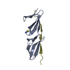
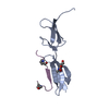
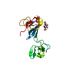
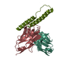
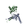
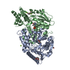
登録情報 データベース : PDB / ID : 3mqlタイトル Crystal structure of the fibronectin 6FnI1-2FnII7FnI fragment Fibronectin 1 キーワード / / / / / 機能・相同性 分子機能 ドメイン・相同性 構成要素
/ / / / / / / / / / / / / / / / / / / / / / / / / / / / / / / / / / / / / / / / / / / / / / / / / / / / / / / / / / / / / / / / / / / / / / / / / / / / / / / / / / / / / / / / / / / / / / / / / / / / / / / / / / / / / / / / / / / / / / / / / / / 生物種 Homo sapiens (ヒト)手法 / / / 解像度 : 3.004 Å データ登録者 Erat, M.C. / Campbell, I.D. / Vakonakis, I. ジャーナル : J.Biol.Chem. / 年 : 2010タイトル : Implications for collagen binding from the crystallographic structure of fibronectin 6FnI1-2FnII7FnI著者 : Erat, M.C. / Schwarz-Linek, U. / Pickford, A.R. / Farndale, R.W. / Campbell, I.D. / Vakonakis, I. 履歴 登録 2010年4月28日 登録サイト / 処理サイト 改定 1.0 2010年8月25日 Provider / タイプ 改定 1.1 2011年7月13日 Group 改定 1.2 2020年7月29日 Group Data collection / Database references ... Data collection / Database references / Derived calculations / Structure summary カテゴリ chem_comp / entity ... chem_comp / entity / pdbx_chem_comp_identifier / pdbx_entity_nonpoly / struct_conn / struct_ref_seq_dif / struct_site / struct_site_gen Item _chem_comp.name / _chem_comp.type ... _chem_comp.name / _chem_comp.type / _entity.pdbx_description / _pdbx_entity_nonpoly.name / _struct_conn.pdbx_leaving_atom_flag / _struct_conn.pdbx_role / _struct_ref_seq_dif.details 解説 / Provider / タイプ 改定 1.3 2023年11月1日 Group Data collection / Database references ... Data collection / Database references / Refinement description / Structure summary カテゴリ chem_comp / chem_comp_atom ... chem_comp / chem_comp_atom / chem_comp_bond / database_2 / pdbx_initial_refinement_model Item / _database_2.pdbx_DOI / _database_2.pdbx_database_accession改定 1.4 2024年10月30日 Group カテゴリ / pdbx_modification_feature
すべて表示 表示を減らす
 ムービー
ムービー コントローラー


 データを開く
 基本情報
基本情報 要素
要素 キーワード
キーワード 機能・相同性情報
 Homo sapiens (ヒト)
 X線回折 /
  データ登録者
 引用
引用 構造の表示
構造の表示 ダウンロードとリンク
 その他のダウンロード
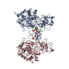


 PDBj
PDBj


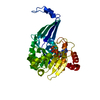
 集合体
集合体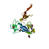
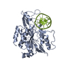
 Pichia pastoris (菌類) / 株 (発現宿主): X33 / 参照: UniProt: B7ZLF0, UniProt: P02751*PLUS
 タイプ: D-saccharide, beta linking / 分子量: 221.208 Da / 分子数: 1 / 由来タイプ: 組換発現 / 式: C8H15NO6
 分子量: 118.174 Da / 分子数: 1 / 由来タイプ: 合成 / 式: C6H14O2 / コメント: 沈殿剤*YM
 分子量: 118.174 Da / 分子数: 2 / 由来タイプ: 合成 / 式: C6H14O2 / コメント: 沈殿剤*YM
 分子量: 18.015 Da / 分子数: 2 / 由来タイプ: 天然 / 式: H2O
 試料調製
試料調製 / ビームライン: X06DA / 波長: 1 Å
 解析
解析