 Movie
Movie Controller

[English] 日本語
 Yorodumi
- PDB-2big: Radiation damage of the Schiff base in phosphoserine aminotransfe... -
+ Open data

- Basic information
| Entry | Database: PDB / ID: 2big | ||||||
|---|---|---|---|---|---|---|---|
| Title | Radiation damage of the Schiff base in phosphoserine aminotransferase (structure I) | ||||||
 Components Components | PHOSPHOSERINE AMINOTRANSFERASE | ||||||
 Keywords Keywords | TRANSFERASE / AMINOTRANSFERASE / PYRIDOXAL-5'-PHOSPHATE / RADIATION DAMAGE | ||||||
| Function / homology |  Function and homology information Function and homology informationphosphoserine transaminase / O-phospho-L-serine:2-oxoglutarate transaminase activity / L-serine biosynthetic process / pyridoxal phosphate binding / cytoplasm Similarity search - Function | ||||||
| Biological species |  BACILLUS ALCALOPHILUS (bacteria) BACILLUS ALCALOPHILUS (bacteria) | ||||||
| Method |  X-RAY DIFFRACTION / SYNCHROTRON / OTHER / Resolution: 1.3 Å X-RAY DIFFRACTION / SYNCHROTRON / OTHER / Resolution: 1.3 Å | ||||||
 Authors Authors | Dubnovitsky, A.P. / Ravelli, R.B.G. / Popov, A.N. / Papageorgiou, A.C. | ||||||
 Citation Citation | Journal: Protein Sci. / Year: 2005 Title: Strain Relief at the Active Site of Phosphoserine Aminotransferase Induced by Radiation Damage. Authors: Dubnovitsky, A.P. / Ravelli, R.B.G. / Popov, A.N. / Papageorgiou, A.C. | ||||||
| History |
|
- Structure visualization
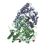
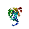
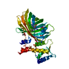
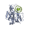
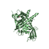
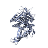
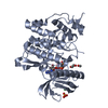
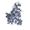
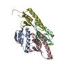
| Structure viewer | Molecule: MolmilJmol/JSmol |
|---|
- Downloads & links
-Download
| PDBx/mmCIF format | 2big.cif.gz | 329.7 KB | Display | PDBx/mmCIF format |
|---|---|---|---|---|
| PDB format | pdb2big.ent.gz | 268.4 KB | Display | PDB format |
| PDBx/mmJSON format | 2big.json.gz | Tree view | PDBx/mmJSON format | |
| Others |  Other downloads Other downloads |
-Validation report
| Arichive directory | https://data.pdbj.org/pub/pdb/validation_reports/bi/2bigftp://data.pdbj.org/pub/pdb/validation_reports/bi/2big | HTTPS FTP |
|---|
-Related structure data
| Related structure data |  2bhxC 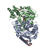 2bi1C  2bi2C  2bi3C  2bi5C  2bi9C  2biaC 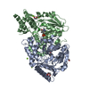 2bieC C: citing same article ( |
|---|---|
| Similar structure data |

-Links
 PDBj
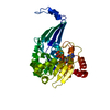
PDBj- Assembly
| Deposited unit | 
| ||||||||
|---|---|---|---|---|---|---|---|---|---|
| 1 |
| ||||||||
| Unit cell |
|
-Components
-Protein , 1 types, 2 molecules AB
| #1: Protein | Mass: 40374.754 Da / Num. of mol.: 2 Source method: isolated from a genetically manipulated source Details: PYRIDOXAL-5'-PHOSPHATE LINKED TO 196 / Source: (gene. exp.) BACILLUS ALCALOPHILUS (bacteria) / Plasmid: PBALC-PSAT / Production host: |
|---|
-Non-polymers , 6 types, 890 molecules 


| #2: Chemical |  Mass: 247.142 Da / Num. of mol.: 2 / Source method: obtained synthetically / Formula: C8H10NO6P Mass: 247.142 Da / Num. of mol.: 2 / Source method: obtained synthetically / Formula: C8H10NO6P#3: Chemical | ChemComp-MG /  Mass: 24.305 Da / Num. of mol.: 5 / Source method: obtained synthetically / Formula: Mg Mass: 24.305 Da / Num. of mol.: 5 / Source method: obtained synthetically / Formula: Mg#4: Chemical | ChemComp-CL /  Mass: 35.453 Da / Num. of mol.: 4 / Source method: obtained synthetically / Formula: Cl Mass: 35.453 Da / Num. of mol.: 4 / Source method: obtained synthetically / Formula: Cl#5: Chemical | ChemComp-PGE / |  Mass: 150.173 Da / Num. of mol.: 1 / Source method: obtained synthetically / Formula: C6H14O4 Mass: 150.173 Da / Num. of mol.: 1 / Source method: obtained synthetically / Formula: C6H14O4#6: Chemical |  Mass: 106.120 Da / Num. of mol.: 2 / Source method: obtained synthetically / Formula: C4H10O3 Mass: 106.120 Da / Num. of mol.: 2 / Source method: obtained synthetically / Formula: C4H10O3#7: Water | ChemComp-HOH / | Mass: 18.015 Da / Num. of mol.: 876 / Source method: isolated from a natural source / Formula: H2O |
|---|
-Details
| Compound details | THIS ENTRY CONTAINS ONE OF NINE STRUCTURES THAT ARE DESCRIBED IN THE ACCOMPANYING JOURNAL ARTICLE. ...THIS ENTRY CONTAINS ONE OF NINE STRUCTURES |
|---|---|
| Has protein modification | N |
-Experimental details
-Experiment
| Experiment | Method: X-RAY DIFFRACTION / Number of used crystals: 1 |
|---|
- Sample preparation
| Crystal | Density Matthews: 2.18 Å3/Da / Density % sol: 43.6 % |
|---|---|
| Crystal grow | pH: 8.2 Details: 30% PEG 400, 200 MM MGCL2, 5% GLYCEROL, 100 MM TRIS PH 8.2 |
-Data collection
| Diffraction | Mean temperature: 100 K |
|---|---|
| Diffraction source | Source: SYNCHROTRON / Site: EMBL/DESY, HAMBURG  / Beamline: BW7A / Wavelength: 0.921 / Beamline: BW7A / Wavelength: 0.921 |
| Detector | Type: MARRESEARCH / Detector: CCD / Date: Jul 12, 2004 |
| Radiation | Protocol: SINGLE WAVELENGTH / Monochromatic (M) / Laue (L): M / Scattering type: x-ray |
| Radiation wavelength | Wavelength: 0.921 Å / Relative weight: 1 |
| Reflection | Resolution: 1.3→12 Å / Num. obs: 187887 / % possible obs: 93.7 % / Observed criterion σ(I): 3 / Redundancy: 6.3 % / Rmerge(I) obs: 0.04 / Net I/σ(I): 96 |
| Reflection shell | Resolution: 1.3→1.33 Å / Rmerge(I) obs: 0.26 / Mean I/σ(I) obs: 3.6 / % possible all: 94.7 |
- Processing
| Software |
| |||||||||||||||||||||||||||||||||
|---|---|---|---|---|---|---|---|---|---|---|---|---|---|---|---|---|---|---|---|---|---|---|---|---|---|---|---|---|---|---|---|---|---|---|
| Refinement | Method to determine structure: OTHER / Resolution: 1.3→12 Å / Num. parameters: 59718 / Num. restraintsaints: 72392 / Cross valid method: FREE R-VALUE / σ(F): 0 / Stereochemistry target values: ENGH AND HUBER Details: ANISOUTROPIC REFINEMENT REDUCED FREE R (NO CUTOFF) BY 0.035. RESIDUES A 215, A 216, A 218, AND B 218 WERE MODELLED AS ALANINES. RESIDUES A 1, A 2, B 1, B 2, B 214, B215 AND B 216 WERE NOT MODELLED.
| |||||||||||||||||||||||||||||||||
| Refine analyze | Num. disordered residues: 31 / Occupancy sum hydrogen: 4936 / Occupancy sum non hydrogen: 6524 | |||||||||||||||||||||||||||||||||
| Refinement step | Cycle: LAST / Resolution: 1.3→12 Å
| |||||||||||||||||||||||||||||||||
| Refine LS restraints |
|
