 Movie
Movie Controller

[English] 日本語
 Yorodumi
- PDB-3mno: Crystal structure of the agonist form of mouse glucocorticoid rec... -
+ Open data

- Basic information
| Entry | Database: PDB / ID: 3mno | ||||||
|---|---|---|---|---|---|---|---|
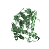
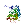
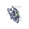
| Title | Crystal structure of the agonist form of mouse glucocorticoid receptor stabilized by (A611V, F608S) mutations at 1.55A | ||||||
 Components Components |
| ||||||
 Keywords Keywords | HORMONE RECEPTOR / protein-ligand complex / steroid nuclear receptor / mouse GR / agonist / co-activator | ||||||
| Function / homology |  Function and homology information Function and homology informationActivated PKN1 stimulates transcription of AR (androgen receptor) regulated genes KLK2 and KLK3 / nuclear receptor-mediated corticosteroid signaling pathway / negative regulation of behavioral fear response / SUMOylation of intracellular receptors / Recycling of bile acids and salts / Synthesis of bile acids and bile salts / Nuclear Receptor transcription pathway / Synthesis of bile acids and bile salts via 7alpha-hydroxycholesterol / Synthesis of bile acids and bile salts via 27-hydroxycholesterol / Endogenous sterols ...Activated PKN1 stimulates transcription of AR (androgen receptor) regulated genes KLK2 and KLK3 / nuclear receptor-mediated corticosteroid signaling pathway / negative regulation of behavioral fear response / SUMOylation of intracellular receptors / Recycling of bile acids and salts / Synthesis of bile acids and bile salts / Nuclear Receptor transcription pathway / Synthesis of bile acids and bile salts via 7alpha-hydroxycholesterol / Synthesis of bile acids and bile salts via 27-hydroxycholesterol / Endogenous sterols / negative regulation of synaptic plasticity / HATs acetylate histones / HSP90 chaperone cycle for steroid hormone receptors (SHR) in the presence of ligand / nuclear receptor-mediated glucocorticoid signaling pathway / positive regulation of nuclear receptor-mediated glucocorticoid signaling pathway / negative regulation of nuclear receptor-mediated glucocorticoid signaling pathway / negative regulation of long-term synaptic depression / regulation of glucocorticoid biosynthetic process / nuclear glucocorticoid receptor activity / Regulation of lipid metabolism by PPARalpha / positive regulation of cell growth involved in cardiac muscle cell development / steroid hormone binding / Cytoprotection by HMOX1 / glucocorticoid metabolic process / neuroinflammatory response / Estrogen-dependent gene expression / response to cortisol / Leydig cell differentiation / nuclear glucocorticoid receptor binding / mammary gland duct morphogenesis / microglia differentiation / nuclear retinoic acid receptor binding / astrocyte differentiation / response to arsenic-containing substance / maternal behavior / positive regulation of female receptivity / negative regulation of vascular permeability / nuclear thyroid hormone receptor binding / RNA polymerase II intronic transcription regulatory region sequence-specific DNA binding / regulation of gluconeogenesis / adrenal gland development / cellular response to glucocorticoid stimulus / cellular response to steroid hormone stimulus / positive regulation of glutamate secretion / response to corticosterone / locomotor rhythm / positive regulation of dendritic spine development / aryl hydrocarbon receptor binding / motor behavior / cellular response to Thyroglobulin triiodothyronine / hormone binding / regulation of glucose metabolic process / regulation of lipid metabolic process / nuclear receptor-mediated steroid hormone signaling pathway / estrogen response element binding / associative learning / cellular response to dexamethasone stimulus / cellular response to transforming growth factor beta stimulus / nuclear retinoid X receptor binding / core promoter sequence-specific DNA binding / transcription regulator inhibitor activity / postsynaptic density, intracellular component / cellular response to hormone stimulus / steroid binding / heat shock protein binding / peroxisome proliferator activated receptor signaling pathway / response to progesterone / DNA polymerase binding / regulation of cellular response to insulin stimulus / positive regulation of adipose tissue development / Hsp70 protein binding / TBP-class protein binding / transcription initiation-coupled chromatin remodeling / nuclear estrogen receptor binding / nuclear receptor binding / positive regulation of cytokine production / RNA polymerase II transcription regulatory region sequence-specific DNA binding / circadian regulation of gene expression / negative regulation of smoothened signaling pathway / promoter-specific chromatin binding / synaptic transmission, glutamatergic / circadian rhythm / Hsp90 protein binding / receptor tyrosine kinase binding / response to wounding / positive regulation of miRNA transcription / DNA-binding transcription repressor activity, RNA polymerase II-specific / mRNA transcription by RNA polymerase II / nuclear receptor activity / RNA polymerase II transcription regulator complex / spindle / sequence-specific double-stranded DNA binding / positive regulation of neuron apoptotic process / regulation of cell population proliferation / regulation of gene expression / double-stranded DNA binding / DNA-binding transcription activator activity, RNA polymerase II-specific / transcription regulator complex / sequence-specific DNA binding / gene expression Similarity search - Function | ||||||
| Biological species |  | ||||||
| Method |  X-RAY DIFFRACTION / SYNCHROTRON / MOLECULAR REPLACEMENT / Resolution: 1.55 Å X-RAY DIFFRACTION / SYNCHROTRON / MOLECULAR REPLACEMENT / Resolution: 1.55 Å | ||||||
 Authors Authors | Schoch, G.A. / Seitz, T. / Benz, J. / Banner, D. / Stihle, M. / D'Arcy, B. / Thoma, R. / Sterner, R. / Hennig, M. / Ruf, A. | ||||||
 Citation Citation | Journal: J.Mol.Biol. / Year: 2010 Title: Enhancing the stability and solubility of the glucocorticoid receptor ligand-binding domain by high-throughput library screening. Authors: Seitz, T. / Thoma, R. / Schoch, G.A. / Stihle, M. / Benz, J. / D'Arcy, B. / Wiget, A. / Ruf, A. / Hennig, M. / Sterner, R. | ||||||
| History |
|
- Structure visualization
| Structure viewer | Molecule: MolmilJmol/JSmol |
|---|
- Downloads & links
-Download
| PDBx/mmCIF format | 3mno.cif.gz | 80 KB | Display | PDBx/mmCIF format |
|---|---|---|---|---|
| PDB format | pdb3mno.ent.gz | 58.7 KB | Display | PDB format |
| PDBx/mmJSON format | 3mno.json.gz | Tree view | PDBx/mmJSON format | |
| Others |  Other downloads Other downloads |
-Validation report
| Arichive directory | https://data.pdbj.org/pub/pdb/validation_reports/mn/3mnoftp://data.pdbj.org/pub/pdb/validation_reports/mn/3mno | HTTPS FTP |
|---|
-Related structure data
| Related structure data | 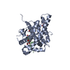 3mneSC  3mnpC S: Starting model for refinement C: citing same article ( |
|---|---|
| Similar structure data |


-Links
 PDBj
PDBj


- Assembly
| Deposited unit | 
| ||||||||
|---|---|---|---|---|---|---|---|---|---|
| 1 |
| ||||||||
| Unit cell |
|
-Components
-Protein / Protein/peptide , 2 types, 2 molecules AB
| #1: Protein | Mass: 30124.143 Da / Num. of mol.: 1 / Fragment: UNP residues 527-783 / Mutation: A611V, F608S Source method: isolated from a genetically manipulated source Source: (gene. exp.)  |
|---|---|
| #2: Protein/peptide | Mass: 1593.844 Da / Num. of mol.: 1 / Fragment: TIF2 coactivator motif, residues 740-752 / Source method: obtained synthetically Details: The sequence occurs naturally in Mus musculus (mouse) References: UniProt: Q61026 |
-Non-polymers , 4 types, 370 molecules 


| #3: Chemical |  Mass: 92.094 Da / Num. of mol.: 2 / Source method: obtained synthetically / Formula: C3H8O3 Mass: 92.094 Da / Num. of mol.: 2 / Source method: obtained synthetically / Formula: C3H8O3#4: Chemical | ChemComp-DEX / |  Mass: 392.461 Da / Num. of mol.: 1 / Source method: obtained synthetically / Formula: C22H29FO5 Mass: 392.461 Da / Num. of mol.: 1 / Source method: obtained synthetically / Formula: C22H29FO5#5: Chemical |  Mass: 58.082 Da / Num. of mol.: 2 / Source method: obtained synthetically / Formula: CNS Mass: 58.082 Da / Num. of mol.: 2 / Source method: obtained synthetically / Formula: CNS#6: Water | ChemComp-HOH / | Mass: 18.015 Da / Num. of mol.: 365 / Source method: isolated from a natural source / Formula: H2O |
|---|
-Experimental details
-Experiment
| Experiment | Method: X-RAY DIFFRACTION / Number of used crystals: 1 |
|---|
- Sample preparation
| Crystal | Density Matthews: 2.99 Å3/Da / Density % sol: 58.92 % |
|---|---|
| Crystal grow | Method: vapor diffusion, hanging drop / pH: 6.5 Details: 0.2 M sodium thiocyanate, 0.1 M Bis-Tris pH 6.5, 0.7 M lithium sulfate, 10 % Glycerol, VAPOR DIFFUSION, HANGING DROP |
-Data collection
| Diffraction | Mean temperature: 100 K |
|---|---|
| Diffraction source | Source: SYNCHROTRON / Site: SLS  / Beamline: X10SA / Wavelength: 1 Å / Beamline: X10SA / Wavelength: 1 Å |
| Detector | Date: Nov 18, 2008 |
| Radiation | Protocol: SINGLE WAVELENGTH / Monochromatic (M) / Laue (L): M / Scattering type: x-ray |
| Radiation wavelength | Wavelength: 1 Å / Relative weight: 1 |
| Reflection | Resolution: 1.55→35.7 Å / Num. all: 53304 / Num. obs: 53304 / % possible obs: 99.7 % / Redundancy: 11.3 % / Biso Wilson estimate: 23.767 Å2 / Rsym value: 0.067 / Net I/σ(I): 21.7 |
| Reflection shell | Resolution: 1.55→1.63 Å / Redundancy: 11.2 % / Mean I/σ(I) obs: 2.9 / Num. unique all: 7760 / Rsym value: 0.846 / % possible all: 100 |
- Processing
| Software |
| ||||||||||||||||||||||||||||||||||||||||||||||||||
|---|---|---|---|---|---|---|---|---|---|---|---|---|---|---|---|---|---|---|---|---|---|---|---|---|---|---|---|---|---|---|---|---|---|---|---|---|---|---|---|---|---|---|---|---|---|---|---|---|---|---|---|
| Refinement | Method to determine structure: MOLECULAR REPLACEMENT Starting model: 3MNE Resolution: 1.55→35.69 Å / Cross valid method: THROUGHOUT / σ(F): 0
| ||||||||||||||||||||||||||||||||||||||||||||||||||
| Displacement parameters | Biso mean: 30.65 Å2
| ||||||||||||||||||||||||||||||||||||||||||||||||||
| Refinement step | Cycle: LAST / Resolution: 1.55→35.69 Å
| ||||||||||||||||||||||||||||||||||||||||||||||||||
| Refine LS restraints |
| ||||||||||||||||||||||||||||||||||||||||||||||||||
| LS refinement shell | Resolution: 1.55→1.64 Å / Total num. of bins used: 9
|
