 Movie
Movie Controller


+ Open data

- Basic information
| Entry | Database: PDB / ID: 2h42 | ||||||
|---|---|---|---|---|---|---|---|
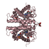
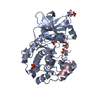
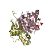
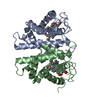
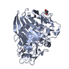
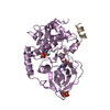
| Title | Crystal structure of PDE5 in complex with sildenafil | ||||||
 Components Components | cGMP-specific 3',5'-cyclic phosphodiesterase | ||||||
 Keywords Keywords | HYDROLASE / viagra / PDE5 | ||||||
| Function / homology |  Function and homology information Function and homology information3',5'-cyclic-GMP phosphodiesterase / RHOBTB1 GTPase cycle / cGMP catabolic process / cGMP effects / 3',5'-cyclic-nucleotide phosphodiesterase activity / cGMP binding / 3',5'-cyclic-GMP phosphodiesterase activity / 3',5'-cyclic-AMP phosphodiesterase activity / Smooth Muscle Contraction / negative regulation of cAMP/PKA signal transduction ...3',5'-cyclic-GMP phosphodiesterase / RHOBTB1 GTPase cycle / cGMP catabolic process / cGMP effects / 3',5'-cyclic-nucleotide phosphodiesterase activity / cGMP binding / 3',5'-cyclic-GMP phosphodiesterase activity / 3',5'-cyclic-AMP phosphodiesterase activity / Smooth Muscle Contraction / negative regulation of cAMP/PKA signal transduction / signal transduction / metal ion binding / cytosol Similarity search - Function | ||||||
| Biological species |  Homo sapiens (human) Homo sapiens (human) | ||||||
| Method |  X-RAY DIFFRACTION / SYNCHROTRON / MOLECULAR REPLACEMENT / Resolution: 2.3 Å X-RAY DIFFRACTION / SYNCHROTRON / MOLECULAR REPLACEMENT / Resolution: 2.3 Å | ||||||
 Authors Authors | Wang, H. / Ke, H. | ||||||
 Citation Citation | Journal: J.Biol.Chem. / Year: 2006 Title: Multiple Conformations of Phosphodiesterase-5: Implications for enzyme function and drug development Authors: Wang, H. / Liu, Y. / Huai, Q. / Cai, J. / Zoraghi, R. / Francis, S.H. / Corbin, J.D. / Robinson, H. / Xin, Z. / Lin, G. / Ke, H. | ||||||
| History |
|
- Structure visualization
| Structure viewer | Molecule: MolmilJmol/JSmol |
|---|
- Downloads & links
-Download
| PDBx/mmCIF format | 2h42.cif.gz | 208.4 KB | Display | PDBx/mmCIF format |
|---|---|---|---|---|
| PDB format | pdb2h42.ent.gz | 168.6 KB | Display | PDB format |
| PDBx/mmJSON format | 2h42.json.gz | Tree view | PDBx/mmJSON format | |
| Others |  Other downloads Other downloads |
-Validation report
| Arichive directory | https://data.pdbj.org/pub/pdb/validation_reports/h4/2h42ftp://data.pdbj.org/pub/pdb/validation_reports/h4/2h42 | HTTPS FTP |
|---|
-Related structure data
| Related structure data |  2h40C 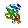 2h44C  1rkpS C: citing same article ( S: Starting model for refinement |
|---|---|
| Similar structure data |


-Links
 PDBj
PDBj


- Assembly
| Deposited unit | 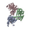
| ||||||||
|---|---|---|---|---|---|---|---|---|---|
| 1 | 
| ||||||||
| 2 | 
| ||||||||
| 3 | 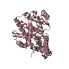
| ||||||||
| 4 | 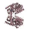
| ||||||||
| Unit cell |
| ||||||||
| Details | Biological unit may be a monomer. However, the crystallographic asymmetric unit contains 3 molecules. |
-Components
| #1: Protein | Mass: 37788.531 Da / Num. of mol.: 3 / Fragment: catalytic domain, residues 535-860 Source method: isolated from a genetically manipulated source Source: (gene. exp.) Homo sapiens (human) / Gene: PDE5A, PDE5 / Plasmid: pET15b / Species (production host): Escherichia coli / Production host:  References: UniProt: O76074, 3',5'-cyclic-GMP phosphodiesterase #2: Chemical |   Mass: 65.409 Da / Num. of mol.: 3 / Source method: obtained synthetically / Formula: Zn Mass: 65.409 Da / Num. of mol.: 3 / Source method: obtained synthetically / Formula: Zn#3: Chemical |   Mass: 24.305 Da / Num. of mol.: 3 / Source method: obtained synthetically / Formula: Mg Mass: 24.305 Da / Num. of mol.: 3 / Source method: obtained synthetically / Formula: Mg#4: Chemical |   Mass: 474.576 Da / Num. of mol.: 3 / Source method: obtained synthetically / Formula: C22H30N6O4S Mass: 474.576 Da / Num. of mol.: 3 / Source method: obtained synthetically / Formula: C22H30N6O4S#5: Water | ChemComp-HOH / |  Mass: 18.015 Da / Num. of mol.: 311 / Source method: isolated from a natural source / Formula: H2O Mass: 18.015 Da / Num. of mol.: 311 / Source method: isolated from a natural source / Formula: H2O |
|---|
-Experimental details
-Experiment
| Experiment | Method: X-RAY DIFFRACTION / Number of used crystals: 1 |
|---|
- Sample preparation
| Crystal | Density Matthews: 3.33 Å3/Da / Density % sol: 63.05 % |
|---|---|
| Crystal grow | Temperature: 277 K / Method: vapor diffusion, hanging drop / pH: 7.5 Details: 1 mM sildenafil mixed with 15 mg/mL PDE5A1 overnight, crystallized against a well buffer of 1.0 M sodium citrate, 2.5% ethanol, 0.1 M HEPES, pH 7.5, VAPOR DIFFUSION, HANGING DROP, temperature 277K |
-Data collection
| Diffraction | Mean temperature: 100 K |
|---|---|
| Diffraction source | Source: SYNCHROTRON / Site: NSLS  / Beamline: X29A / Wavelength: 1.1 Å / Beamline: X29A / Wavelength: 1.1 Å |
| Detector | Type: ADSC QUANTUM 315 / Detector: CCD / Date: Nov 19, 2003 |
| Radiation | Protocol: SINGLE WAVELENGTH / Monochromatic (M) / Laue (L): M / Scattering type: x-ray |
| Radiation wavelength | Wavelength: 1.1 Å / Relative weight: 1 |
| Reflection | Resolution: 2.3→30 Å / Num. obs: 68786 / % possible obs: 99.9 % / Observed criterion σ(F): 0 / Observed criterion σ(I): 0 / Redundancy: 16 % / Rmerge(I) obs: 0.117 / Net I/σ(I): 6.7 |
| Reflection shell | Resolution: 2.3→2.4 Å / Rmerge(I) obs: 0.58 / Mean I/σ(I) obs: 4.8 / % possible all: 100 |
- Processing
| Software |
| ||||||||||||||||||
|---|---|---|---|---|---|---|---|---|---|---|---|---|---|---|---|---|---|---|---|
| Refinement | Method to determine structure: MOLECULAR REPLACEMENT Starting model: PDB ENTRY 1RKP Resolution: 2.3→30 Å / σ(F): 0 / σ(I): 0 / Stereochemistry target values: Engh & Huber
| ||||||||||||||||||
| Refinement step | Cycle: LAST / Resolution: 2.3→30 Å
| ||||||||||||||||||
| Refine LS restraints |
| ||||||||||||||||||
| Xplor file |
|
