 Movie
Movie Controller


+ Open data

- Basic information
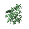
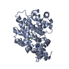
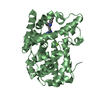
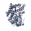
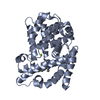
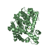
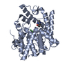
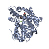
| Entry | Database: PDB / ID: 2ouq | |||||||||
|---|---|---|---|---|---|---|---|---|---|---|
| Title | crystal structure of PDE10A2 in complex with GMP | |||||||||
 Components Components | cAMP and cAMP-inhibited cGMP 3',5'-cyclic phosphodiesterase 10A | |||||||||
 Keywords Keywords | HYDROLASE / pde / gmp complex | |||||||||
| Function / homology |  Function and homology information Function and homology information3',5'-cGMP-stimulated cyclic-nucleotide phosphodiesterase activity / negative regulation of receptor guanylyl cyclase signaling pathway / 3',5'-cyclic-nucleotide phosphodiesterase / cGMP catabolic process / cAMP catabolic process / cGMP effects / 3',5'-cyclic-nucleotide phosphodiesterase activity / cGMP binding / 3',5'-cyclic-GMP phosphodiesterase activity / 3',5'-cyclic-AMP phosphodiesterase activity ...3',5'-cGMP-stimulated cyclic-nucleotide phosphodiesterase activity / negative regulation of receptor guanylyl cyclase signaling pathway / 3',5'-cyclic-nucleotide phosphodiesterase / cGMP catabolic process / cAMP catabolic process / cGMP effects / 3',5'-cyclic-nucleotide phosphodiesterase activity / cGMP binding / 3',5'-cyclic-GMP phosphodiesterase activity / 3',5'-cyclic-AMP phosphodiesterase activity / regulation of adenylate cyclase-activating G protein-coupled receptor signaling pathway / cAMP binding / negative regulation of cAMP/PKA signal transduction / G alpha (s) signalling events / glutamatergic synapse / signal transduction / metal ion binding / cytosol Similarity search - Function | |||||||||
| Biological species |  Homo sapiens (human) Homo sapiens (human) | |||||||||
| Method |  X-RAY DIFFRACTION / SYNCHROTRON / MOLECULAR REPLACEMENT / Resolution: 1.9 Å X-RAY DIFFRACTION / SYNCHROTRON / MOLECULAR REPLACEMENT / Resolution: 1.9 Å | |||||||||
 Authors Authors | Wang, H.C. / Liu, Y.D. / Hou, J. / Zheng, M.Y. / Robinson, H. | |||||||||
 Citation Citation | Journal: Proc.Natl.Acad.Sci.Usa / Year: 2007 Title: From the Cover: Structural insight into substrate specificity of phosphodiesterase 10. Authors: Wang, H. / Liu, Y. / Hou, J. / Zheng, M. / Robinson, H. / Ke, H. | |||||||||
| History |
|
- Structure visualization
| Structure viewer | Molecule: MolmilJmol/JSmol |
|---|
- Downloads & links
-Download
| PDBx/mmCIF format | 2ouq.cif.gz | 146.7 KB | Display | PDBx/mmCIF format |
|---|---|---|---|---|
| PDB format | pdb2ouq.ent.gz | 114.2 KB | Display | PDB format |
| PDBx/mmJSON format | 2ouq.json.gz | Tree view | PDBx/mmJSON format | |
| Others |  Other downloads Other downloads |
-Validation report
| Arichive directory | https://data.pdbj.org/pub/pdb/validation_reports/ou/2ouqftp://data.pdbj.org/pub/pdb/validation_reports/ou/2ouq | HTTPS FTP |
|---|
-Related structure data
| Related structure data |  2ounC 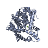 2oupC  2ourC  2ousC 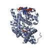 2ouuC  2ouvC  2ouyC C: citing same article ( |
|---|---|
| Similar structure data |


-Links
 PDBj
PDBj


- Assembly
| Deposited unit | 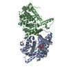
| ||||||||
|---|---|---|---|---|---|---|---|---|---|
| 1 | 
| ||||||||
| 2 | 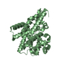
| ||||||||
| Unit cell |
|
-Components
| #1: Protein | Mass: 38306.078 Da / Num. of mol.: 2 / Fragment: catalytic domain Source method: isolated from a genetically manipulated source Source: (gene. exp.) Homo sapiens (human) / Strain: pde10a2 / Gene: PDE10A / Plasmid: pET15b / Species (production host): Escherichia coli / Production host:  References: UniProt: Q9Y233, 3',5'-cyclic-nucleotide phosphodiesterase #2: Chemical |   Mass: 65.409 Da / Num. of mol.: 2 / Source method: obtained synthetically / Formula: Zn Mass: 65.409 Da / Num. of mol.: 2 / Source method: obtained synthetically / Formula: Zn#3: Chemical |   Mass: 24.305 Da / Num. of mol.: 2 / Source method: obtained synthetically / Formula: Mg Mass: 24.305 Da / Num. of mol.: 2 / Source method: obtained synthetically / Formula: Mg#4: Chemical | ChemComp-5GP / |   Mass: 363.221 Da / Num. of mol.: 1 / Source method: obtained synthetically / Formula: C10H14N5O8P Mass: 363.221 Da / Num. of mol.: 1 / Source method: obtained synthetically / Formula: C10H14N5O8P#5: Water | ChemComp-HOH / |  Mass: 18.015 Da / Num. of mol.: 324 / Source method: isolated from a natural source / Formula: H2O Mass: 18.015 Da / Num. of mol.: 324 / Source method: isolated from a natural source / Formula: H2OHas protein modification | N | |
|---|
-Experimental details
-Experiment
| Experiment | Method: X-RAY DIFFRACTION / Number of used crystals: 1 |
|---|
- Sample preparation
| Crystal | Density Matthews: 2.11 Å3/Da / Density % sol: 41.65 % |
|---|---|
| Crystal grow | Temperature: 277 K / Method: vapor diffusion, hanging drop / pH: 7.5 Details: soaked the PDE10A2 crystals in 30 mM cGMP in a buffer of 16% PEG8000, 0.1 M HEPES (pH 7.5), 0.1 M MgCl2, 60 mM BME for 1.5 hours, VAPOR DIFFUSION, HANGING DROP, temperature 277K |
-Data collection
| Diffraction | Mean temperature: 100 K |
|---|---|
| Diffraction source | Source: SYNCHROTRON / Site: NSLS  / Beamline: X29A / Wavelength: 1.1 / Beamline: X29A / Wavelength: 1.1 |
| Detector | Type: ADSC QUANTUM 315 / Detector: CCD / Date: Mar 15, 2006 |
| Radiation | Protocol: SINGLE WAVELENGTH / Monochromatic (M) / Laue (L): M / Scattering type: x-ray |
| Radiation wavelength | Wavelength: 1.1 Å / Relative weight: 1 |
| Reflection | Resolution: 1.9→30 Å / Num. obs: 48795 / Observed criterion σ(F): 0 / Observed criterion σ(I): 0 / Redundancy: 6.7 % / Rmerge(I) obs: 0.071 / Net I/σ(I): 7.9 |
- Processing
| Software |
| ||||||||||||||||||
|---|---|---|---|---|---|---|---|---|---|---|---|---|---|---|---|---|---|---|---|
| Refinement | Method to determine structure: MOLECULAR REPLACEMENT Starting model: PDE10A2 native Resolution: 1.9→30 Å / Isotropic thermal model: iostropic / σ(F): 0 / σ(I): 0 / Stereochemistry target values: Engh & Huber
| ||||||||||||||||||
| Displacement parameters | Biso mean: 27.1 Å2 | ||||||||||||||||||
| Refinement step | Cycle: LAST / Resolution: 1.9→30 Å
| ||||||||||||||||||
| Refine LS restraints |
|
