[English] 日本語
Yorodumi
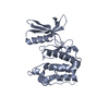
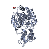
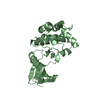
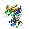
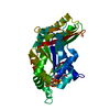
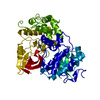
- PDB-3mn3: An inhibited conformation for the protein kinase domain of the Sa... -

+
Open data


ID or keywords:

Loading...

-
Basic information

Entry
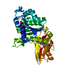
Database: PDB / ID: 3mn3
TitleAn inhibited conformation for the protein kinase domain of the Saccharomyces cerevisiae AMPK homolog Snf1
ComponentsCarbon catabolite-derepressing protein kinase
KeywordsTRANSFERASE / SNF1 / kinase domain / autoinhibitory region
Function / homology
Function and homology information


fungal-type cell wall assembly / positive regulation of pseudohyphal growth / positive regulation of filamentous growth of a population of unicellular organisms in response to starvation / AMPK inhibits chREBP transcriptional activation activity / Energy dependent regulation of mTOR by LKB1-AMPK / single-species surface biofilm formation / regulation of cellular response to glucose starvation / regulation of invasive growth in response to glucose limitation / cellular bud neck septin ring / Carnitine metabolism ...fungal-type cell wall assembly / positive regulation of pseudohyphal growth / positive regulation of filamentous growth of a population of unicellular organisms in response to starvation / AMPK inhibits chREBP transcriptional activation activity / Energy dependent regulation of mTOR by LKB1-AMPK / single-species surface biofilm formation / regulation of cellular response to glucose starvation / regulation of invasive growth in response to glucose limitation / cellular bud neck septin ring / Carnitine metabolism / invasive growth in response to glucose limitation / Macroautophagy / filamentous growth / nucleotide-activated protein kinase complex / vacuolar membrane / AMP-activated protein kinase activity / nuclear envelope lumen / establishment of mitotic spindle orientation / positive regulation of macroautophagy / response to unfolded protein / positive regulation of gluconeogenesis / response to endoplasmic reticulum stress / guanyl-nucleotide exchange factor activity / molecular function activator activity / nuclear membrane / negative regulation of translation / non-specific serine/threonine protein kinase / protein kinase activity / intracellular signal transduction / phosphorylation / protein serine kinase activity / protein serine/threonine kinase activity / mitochondrion / ATP binding / identical protein binding / nucleus / cytoplasm
Similarity search - Function
Carbon catabolite-derepressing protein kinase, ubiquitin-associated domain / Ubiquitin associated domain (UBA) / AMPK, C-terminal adenylate sensor domain / Adenylate sensor of SNF1-like protein kinase / KA1 domain/Ssp2, C-terminal / Transferase(Phosphotransferase) domain 1 / Transferase(Phosphotransferase); domain 1 / Phosphorylase Kinase; domain 1 / Phosphorylase Kinase; domain 1 / Serine/threonine-protein kinase, active site ...Carbon catabolite-derepressing protein kinase, ubiquitin-associated domain / Ubiquitin associated domain (UBA) / AMPK, C-terminal adenylate sensor domain / Adenylate sensor of SNF1-like protein kinase / KA1 domain/Ssp2, C-terminal / Transferase(Phosphotransferase) domain 1 / Transferase(Phosphotransferase); domain 1 / Phosphorylase Kinase; domain 1 / Phosphorylase Kinase; domain 1 / Serine/threonine-protein kinase, active site / Serine/Threonine protein kinases active-site signature. / Protein kinase domain / Serine/Threonine protein kinases, catalytic domain / Protein kinase, ATP binding site / Protein kinases ATP-binding region signature. / Protein kinase domain profile. / Protein kinase domain / Protein kinase-like domain superfamily / 2-Layer Sandwich / Orthogonal Bundle / Mainly Alpha / Alpha Beta
Similarity search - Domain/homology
Carbon catabolite-derepressing protein kinase
Similarity search - Component
Biological speciesSaccharomyces cerevisiae (brewer's yeast)
MethodX-RAY DIFFRACTION / SYNCHROTRON / MOLECULAR REPLACEMENT / Resolution: 2.38 Å
AuthorsRudolph, M.J. / Amodeo, G.A. / Tong, L.
CitationJournal: Acta Crystallogr.,Sect.F / Year: 2010
Title: An inhibited conformation for the protein kinase domain of the Saccharomyces cerevisiae AMPK homolog Snf1.
Authors: Rudolph, M.J. / Amodeo, G.A. / Tong, L.
History
DepositionApr 20, 2010Deposition site: RCSB / Processing site: RCSB
Revision 1.0Sep 15, 2010Provider: repository / Type: Initial release
Revision 1.1Jul 13, 2011Group: Version format compliance
Revision 1.2Feb 21, 2024Group: Data collection / Database references / Category: chem_comp_atom / chem_comp_bond / database_2
Item: _database_2.pdbx_DOI / _database_2.pdbx_database_accession

-
Structure visualization

Structure viewerMolecule:
MolmilJmol/JSmol

Downloads & links

-
Assembly

Deposited unit
A: Carbon catabolite-derepressing protein kinase


Theoretical massNumber of molelcules
Total (without water)31,0281
Polymers31,0281
Non-polymers00
Water97354
1


  • Idetical with deposited unit
  • defined by author
TypeNameSymmetry operationNumber
identity operation1_555x,y,z1
2
A: Carbon catabolite-derepressing protein kinase

A: Carbon catabolite-derepressing protein kinase


Theoretical massNumber of molelcules
Total (without water)62,0562
Polymers62,0562
Non-polymers00
Water362
TypeNameSymmetry operationNumber
identity operation1_555x,y,z1
crystal symmetry operation10_565-x,-y+1,z1
Buried area2610 Å2
ΔGint-19 kcal/mol
Surface area24360 Å2
MethodPISA
Unit cell
Length a, b, c (Å)76.981, 76.981, 286.215
Angle α, β, γ (deg.)90.000, 90.000, 90.000
Int Tables number98
Space group name H-MI4122

-
Components

#1: Protein Carbon catabolite-derepressing protein kinase


Mass: 31028.217 Da / Num. of mol.: 1 / Fragment: UNP residues 50-320
Source method: isolated from a genetically manipulated source
Source: (gene. exp.) Saccharomyces cerevisiae (brewer's yeast)
Gene: SNF1, CAT1, CCR1, GLC2, PAS14, YDR477W, D8035.20 / Plasmid: pET28a / Production host: Escherichia coli (E. coli) / Strain (production host): Bl21(DE3)
References: UniProt: P06782, non-specific serine/threonine protein kinase
#2: Water ChemComp-HOH / water


Mass: 18.015 Da / Num. of mol.: 54 / Source method: isolated from a natural source / Formula: H2O

-
Experimental details

-
Experiment

ExperimentMethod: X-RAY DIFFRACTION / Number of used crystals: 1

-
Sample preparation

CrystalDensity Matthews: 3.42 Å3/Da / Density % sol: 64 %
Crystal growTemperature: 294 K / Method: vapor diffusion / pH: 7.5
Details: 10% (w/v) PEG3350 and 50mM Na2SO4, pH 7.5, VAPOR DIFFUSION, temperature 294K

-
Data collection

DiffractionMean temperature: 100 K
Diffraction sourceSource: SYNCHROTRON / Site: NSLS / Beamline: X4C / Wavelength: 0.9815 Å
DetectorType: MAR scanner 345 mm plate / Detector: IMAGE PLATE / Date: Apr 1, 2009
RadiationMonochromator: horizontally deflecting and focusing crystal preceded by a vertically focusing mirror
Protocol: SINGLE WAVELENGTH / Monochromatic (M) / Laue (L): M / Scattering type: x-ray
Radiation wavelengthWavelength: 0.9815 Å / Relative weight: 1
ReflectionResolution: 2.38→30 Å / Num. all: 19910 / Num. obs: 19799 / % possible obs: 99 % / Observed criterion σ(F): 3 / Observed criterion σ(I): 3
Reflection shellResolution: 2.38→2.48 Å / % possible all: 99

-
Processing

Software
NameVersionClassificationNB
REFMAC5.2.0019refinement
PDB_EXTRACT3.1data extraction
MAR345dtbdata collection
DENZOdata reduction
SCALEPACKdata scaling
COMOphasing
RefinementMethod to determine structure: MOLECULAR REPLACEMENT / Resolution: 2.38→30 Å / Cor.coef. Fo:Fc: 0.938 / Cor.coef. Fo:Fc free: 0.933 / Occupancy max: 1 / Occupancy min: 1 / SU B: 15.093 / SU ML: 0.172 / Cross valid method: THROUGHOUT / σ(F): 0 / ESU R: 0.286 / ESU R Free: 0.215 / Stereochemistry target values: MAXIMUM LIKELIHOOD / Details: HYDROGENS HAVE BEEN ADDED IN THE RIDING POSITIONS
RfactorNum. reflection% reflectionSelection details
Rfree0.246 872 5 %RANDOM
Rwork0.23 ---
obs0.231 17349 98.97 %-
Solvent computationIon probe radii: 0.8 Å / Shrinkage radii: 0.8 Å / VDW probe radii: 1.2 Å / Solvent model: MASK
Displacement parametersBiso max: 89.14 Å2 / Biso mean: 59.94 Å2 / Biso min: 40.67 Å2
Baniso -1Baniso -2Baniso -3
1-2.88 Å20 Å20 Å2
2--2.88 Å20 Å2
3----5.76 Å2
Refinement stepCycle: LAST / Resolution: 2.38→30 Å
ProteinNucleic acidLigandSolventTotal
Num. atoms2040 0 0 54 2094
Refine LS restraints
Refine-IDTypeDev idealDev ideal targetNumber
X-RAY DIFFRACTIONr_bond_refined_d0.010.0222085
X-RAY DIFFRACTIONr_angle_refined_deg1.271.9772819
X-RAY DIFFRACTIONr_dihedral_angle_1_deg5.6055249
X-RAY DIFFRACTIONr_dihedral_angle_2_deg39.14223.87193
X-RAY DIFFRACTIONr_dihedral_angle_3_deg16.7615382
X-RAY DIFFRACTIONr_dihedral_angle_4_deg21.351512
X-RAY DIFFRACTIONr_chiral_restr0.080.2317
X-RAY DIFFRACTIONr_gen_planes_refined0.0040.021544
X-RAY DIFFRACTIONr_nbd_refined0.1960.2858
X-RAY DIFFRACTIONr_nbtor_refined0.3050.21392
X-RAY DIFFRACTIONr_xyhbond_nbd_refined0.130.277
X-RAY DIFFRACTIONr_symmetry_vdw_refined0.2110.230
X-RAY DIFFRACTIONr_symmetry_hbond_refined0.2110.25
X-RAY DIFFRACTIONr_mcbond_it0.6151.51304
X-RAY DIFFRACTIONr_mcangle_it1.13322044
X-RAY DIFFRACTIONr_scbond_it1.2933892
X-RAY DIFFRACTIONr_scangle_it2.1054.5775
LS refinement shellResolution: 2.38→2.457 Å / Total num. of bins used: 20
RfactorNum. reflection% reflection
Rfree0.346 64 -
Rwork0.3 1139 -
all-1203 -
obs--96.09 %
Refinement TLS params.

Method: refined / Refine-ID: X-RAY DIFFRACTION

IDL112)L122)L132)L222)L232)L332)S11 (Å °)S12 (Å °)S13 (Å °)S21 (Å °)S22 (Å °)S23 (Å °)S31 (Å °)S32 (Å °)S33 (Å °)T112)T122)T132)T222)T232)T332)Origin x (Å)Origin y (Å)Origin z (Å)
13.8466-1.4269-0.70633.07320.40835.1587-0.08970.05040.45280.09880.26110.2069-0.5944-0.6458-0.1714-0.04280.05920.0172-0.06250.0434-0.1016-18.01733.836231.712
21.34730.3817-0.40182.3262-0.18772.7232-0.14880.13370.0294-0.02070.1864-0.15160.09410.0266-0.0376-0.1036-0.1521-0.038-0.0514-0.0326-0.1289-4.343622.2612.4289
Refinement TLS group
IDRefine-IDRefine TLS-IDAuth asym-IDAuth seq-ID
1X-RAY DIFFRACTION1A50 - 135
2X-RAY DIFFRACTION2A136 - 320

+
About Yorodumi

-
News

-
Feb 9, 2022. New format data for meta-information of EMDB entries

New format data for meta-information of EMDB entries

  • Version 3 of the EMDB header file is now the official format.
  • The previous official version 1.9 will be removed from the archive.

Related info.:EMDB header

External links:wwPDB to switch to version 3 of the EMDB data model

-
Aug 12, 2020. Covid-19 info

Covid-19 info

URL: https://pdbjlvh1.pdbj.org/emnavi/covid19.php

New page: Covid-19 featured information page in EM Navigator.

Related info.:Covid-19 info / Mar 5, 2020. Novel coronavirus structure data

+
Mar 5, 2020. Novel coronavirus structure data

Novel coronavirus structure data

Related info.:Yorodumi Speices / Aug 12, 2020. Covid-19 info

External links:COVID-19 featured content - PDBj / Molecule of the Month (242):Coronavirus Proteases

+
Jan 31, 2019. EMDB accession codes are about to change! (news from PDBe EMDB page)

EMDB accession codes are about to change! (news from PDBe EMDB page)

  • The allocation of 4 digits for EMDB accession codes will soon come to an end. Whilst these codes will remain in use, new EMDB accession codes will include an additional digit and will expand incrementally as the available range of codes is exhausted. The current 4-digit format prefixed with “EMD-” (i.e. EMD-XXXX) will advance to a 5-digit format (i.e. EMD-XXXXX), and so on. It is currently estimated that the 4-digit codes will be depleted around Spring 2019, at which point the 5-digit format will come into force.
  • The EM Navigator/Yorodumi systems omit the EMD- prefix.

Related info.:Q: What is EMD? / ID/Accession-code notation in Yorodumi/EM Navigator

External links:EMDB Accession Codes are Changing Soon! / Contact to PDBj

+
Jul 12, 2017. Major update of PDB

Major update of PDB

  • wwPDB released updated PDB data conforming to the new PDBx/mmCIF dictionary.
  • This is a major update changing the version number from 4 to 5, and with Remediation, in which all the entries are updated.
  • In this update, many items about electron microscopy experimental information are reorganized (e.g. em_software).
  • Now, EM Navigator and Yorodumi are based on the updated data.

External links:wwPDB Remediation / Enriched Model Files Conforming to OneDep Data Standards Now Available in the PDB FTP Archive

-
Yorodumi

Thousand views of thousand structures

  • Yorodumi is a browser for structure data from EMDB, PDB, SASBDB, etc.
  • This page is also the successor to EM Navigator detail page, and also detail information page/front-end page for Omokage search.
  • The word "yorodu" (or yorozu) is an old Japanese word meaning "ten thousand". "mi" (miru) is to see.

Related info.:EMDB / PDB / SASBDB / Comparison of 3 databanks / Yorodumi Search / Aug 31, 2016. New EM Navigator & Yorodumi / Yorodumi Papers / Jmol/JSmol / Function and homology information / Changes in new EM Navigator and Yorodumi

Read more