 Movie
Movie Controller


+ Open data

- Basic information
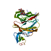
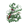
| Entry | Database: PDB / ID: 6s76 | |||||||||
|---|---|---|---|---|---|---|---|---|---|---|
| Title | Crystal structure of human Nek7 | |||||||||
 Components Components | Serine/threonine-protein kinase Nek7 | |||||||||
 Keywords Keywords | CELL CYCLE / Kinase | |||||||||
| Function / homology |  Function and homology information Function and homology informationNEK6-subfamily protein kinase / Activation of NIMA Kinases NEK9, NEK6, NEK7 / Nuclear Pore Complex (NPC) Disassembly / cellular response to potassium ion / positive regulation of NLRP3 inflammasome complex assembly / positive regulation of telomere maintenance / microtubule organizing center / spindle assembly / regulation of mitotic cell cycle / EML4 and NUDC in mitotic spindle formation ...NEK6-subfamily protein kinase / Activation of NIMA Kinases NEK9, NEK6, NEK7 / Nuclear Pore Complex (NPC) Disassembly / cellular response to potassium ion / positive regulation of NLRP3 inflammasome complex assembly / positive regulation of telomere maintenance / microtubule organizing center / spindle assembly / regulation of mitotic cell cycle / EML4 and NUDC in mitotic spindle formation / molecular function activator activity / spindle pole / microtubule / protein phosphorylation / protein serine kinase activity / protein serine/threonine kinase activity / centrosome / nucleoplasm / ATP binding / metal ion binding / nucleus / cytoplasm Similarity search - Function | |||||||||
| Biological species |  Homo sapiens (human) Homo sapiens (human) | |||||||||
| Method |  X-RAY DIFFRACTION / SYNCHROTRON / MOLECULAR REPLACEMENT / Resolution: 3.38 Å X-RAY DIFFRACTION / SYNCHROTRON / MOLECULAR REPLACEMENT / Resolution: 3.38 Å | |||||||||
 Authors Authors | Nasir, N. / Bayliss, R. | |||||||||
| Funding support |  United Kingdom, 2items United Kingdom, 2items
| |||||||||
 Citation Citation | Journal: Biochem.J. / Year: 2020 Title: Nek7 conformational flexibility and inhibitor binding probed through protein engineering of the R-spine. Authors: Byrne, M.J. / Nasir, N. / Basmadjian, C. / Bhatia, C. / Cunnison, R.F. / Carr, K.H. / Mas-Droux, C. / Yeoh, S. / Cano, C. / Bayliss, R. | |||||||||
| History |
|
- Structure visualization
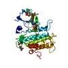
| Structure viewer | Molecule: MolmilJmol/JSmol |
|---|
- Downloads & links
-Download
| PDBx/mmCIF format | 6s76.cif.gz | 414 KB | Display | PDBx/mmCIF format |
|---|---|---|---|---|
| PDB format | pdb6s76.ent.gz | 273 KB | Display | PDB format |
| PDBx/mmJSON format | 6s76.json.gz | Tree view | PDBx/mmJSON format | |
| Others |  Other downloads Other downloads |
-Validation report
| Arichive directory | https://data.pdbj.org/pub/pdb/validation_reports/s7/6s76ftp://data.pdbj.org/pub/pdb/validation_reports/s7/6s76 | HTTPS FTP |
|---|
-Related structure data
| Related structure data | 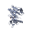 6s73C 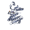 6s75C 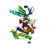 6sk9C  6gt1 C: citing same article ( S: Starting model for refinement |
|---|---|
| Similar structure data |


-Links
 PDBj
PDBj
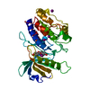
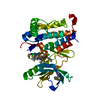
- Assembly
| Deposited unit | 
| ||||||||||||
|---|---|---|---|---|---|---|---|---|---|---|---|---|---|
| 1 | 
| ||||||||||||
| 2 | 
| ||||||||||||
| 3 | 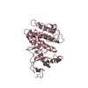
| ||||||||||||
| 4 | 
| ||||||||||||
| Unit cell |
|
-Components
| #1: Protein | Mass: 35670.113 Da / Num. of mol.: 4 Source method: isolated from a genetically manipulated source Source: (gene. exp.) Homo sapiens (human) / Gene: NEK7 / Production host:  References: UniProt: Q8TDX7, non-specific serine/threonine protein kinase #2: Chemical | ChemComp-PEG / |   Mass: 106.120 Da / Num. of mol.: 1 / Source method: obtained synthetically / Formula: C4H10O3 Mass: 106.120 Da / Num. of mol.: 1 / Source method: obtained synthetically / Formula: C4H10O3Has ligand of interest | N | |
|---|
-Experimental details
-Experiment
| Experiment | Method: X-RAY DIFFRACTION / Number of used crystals: 1 |
|---|
- Sample preparation
| Crystal | Density Matthews: 2.89 Å3/Da / Density % sol: 57.4 % |
|---|---|
| Crystal grow | Temperature: 293 K / Method: vapor diffusion, sitting drop / pH: 7 Details: 20 % w/v Polyethylene glycol 3,350, 150 mM di-Sodium DL-malate; pH 7.0 |
-Data collection
| Diffraction | Mean temperature: 80 K / Serial crystal experiment: N |
|---|---|
| Diffraction source | Source: SYNCHROTRON / Site: Diamond / Beamline: I03 / Wavelength: 0.97625 Å |
| Detector | Type: DECTRIS PILATUS3 6M / Detector: PIXEL / Date: Mar 4, 2019 |
| Radiation | Protocol: SINGLE WAVELENGTH / Monochromatic (M) / Laue (L): M / Scattering type: x-ray |
| Radiation wavelength | Wavelength: 0.97625 Å / Relative weight: 1 |
| Reflection | Resolution: 3.38→74.74 Å / Num. obs: 19383 / % possible obs: 99.46 % / Redundancy: 3.3 % / Biso Wilson estimate: 88.34 Å2 / CC1/2: 0.986 / Rmerge(I) obs: 0.101 / Net I/σ(I): 6.54 |
| Reflection shell | Resolution: 3.38→3.5 Å / Num. unique obs: 6623 |
- Processing
| Software |
| ||||||||||||||||||||||||||||||||||||||||||||||||||||||||
|---|---|---|---|---|---|---|---|---|---|---|---|---|---|---|---|---|---|---|---|---|---|---|---|---|---|---|---|---|---|---|---|---|---|---|---|---|---|---|---|---|---|---|---|---|---|---|---|---|---|---|---|---|---|---|---|---|---|
| Refinement | Method to determine structure: MOLECULAR REPLACEMENT Starting model: 6GT1 6gt1 Resolution: 3.38→74.74 Å / SU ML: 0.4454 / Cross valid method: FREE R-VALUE / σ(F): 1.35 / Phase error: 35.5528
| ||||||||||||||||||||||||||||||||||||||||||||||||||||||||
| Solvent computation | Shrinkage radii: 0.9 Å / VDW probe radii: 1.11 Å | ||||||||||||||||||||||||||||||||||||||||||||||||||||||||
| Refinement step | Cycle: LAST / Resolution: 3.38→74.74 Å
| ||||||||||||||||||||||||||||||||||||||||||||||||||||||||
| Refine LS restraints |
| ||||||||||||||||||||||||||||||||||||||||||||||||||||||||
| LS refinement shell |
|
