 Movie
Movie Controller

[English] 日本語
 Yorodumi
- PDB-3md0: Crystal structure of arginine/ornithine transport system ATPase f... -
+ Open data

- Basic information
| Entry | Database: PDB / ID: 3md0 | ||||||
|---|---|---|---|---|---|---|---|
| Title | Crystal structure of arginine/ornithine transport system ATPase from Mycobacterium tuberculosis bound to GDP (a RAS-like GTPase superfamily protein) | ||||||
 Components Components | Arginine/ornithine transport system ATPase | ||||||
 Keywords Keywords | TRANSPORT PROTEIN / Seattle Structural Genomics Center for Infectious Disease / RAS / RAS-like GTPase / allosteric modulator / tuberculosis / ATP-binding / Nucleotide-binding / SSGCID | ||||||
| Function / homology |  Function and homology information Function and homology informationHydrolases; Acting on acid anhydrides / GTPase activity / GTP binding / ATP binding / cytoplasm Similarity search - Function | ||||||
| Biological species |   Mycobacterium tuberculosis (bacteria) Mycobacterium tuberculosis (bacteria) | ||||||
| Method |  X-RAY DIFFRACTION / MOLECULAR REPLACEMENT / Resolution: 2.45 Å X-RAY DIFFRACTION / MOLECULAR REPLACEMENT / Resolution: 2.45 Å | ||||||
 Authors Authors | Seattle Structural Genomics Center for Infectious Disease (SSGCID) | ||||||
 Citation Citation | Journal: J.Struct.Funct.Genom. / Year: 2015 Title: Crystal structures of Mycobacterial MeaB and MMAA-like GTPases. Authors: Edwards, T.E. / Baugh, L. / Bullen, J. / Baydo, R.O. / Witte, P. / Thompkins, K. / Phan, I.Q. / Abendroth, J. / Clifton, M.C. / Sankaran, B. / Van Voorhis, W.C. / Myler, P.J. / Staker, B.L. ...Authors: Edwards, T.E. / Baugh, L. / Bullen, J. / Baydo, R.O. / Witte, P. / Thompkins, K. / Phan, I.Q. / Abendroth, J. / Clifton, M.C. / Sankaran, B. / Van Voorhis, W.C. / Myler, P.J. / Staker, B.L. / Grundner, C. / Lorimer, D.D. #1: Journal: Tuberculosis (Edinb) / Year: 2014Title: Increasing the structural coverage of tuberculosis drug targets. Authors: Baugh, L. / Phan, I. / Begley, D.W. / Clifton, M.C. / Armour, B. / Dranow, D.M. / Taylor, B.M. / Muruthi, M.M. / Abendroth, J. / Fairman, J.W. / Fox, D. / Dieterich, S.H. / Staker, B.L. / ...Authors: Baugh, L. / Phan, I. / Begley, D.W. / Clifton, M.C. / Armour, B. / Dranow, D.M. / Taylor, B.M. / Muruthi, M.M. / Abendroth, J. / Fairman, J.W. / Fox, D. / Dieterich, S.H. / Staker, B.L. / Gardberg, A.S. / Choi, R. / Hewitt, S.N. / Napuli, A.J. / Myers, J. / Barrett, L.K. / Zhang, Y. / Ferrell, M. / Mundt, E. / Thompkins, K. / Tran, N. / Lyons-Abbott, S. / Abramov, A. / Sekar, A. / Serbzhinskiy, D. / Lorimer, D. / Buchko, G.W. / Stacy, R. / Stewart, L.J. / Edwards, T.E. / Van Voorhis, W.C. / Myler, P.J. | ||||||
| History |
|
- Structure visualization
| Structure viewer | Molecule: MolmilJmol/JSmol |
|---|
- Downloads & links
-Download
| PDBx/mmCIF format | 3md0.cif.gz | 132.4 KB | Display | PDBx/mmCIF format |
|---|---|---|---|---|
| PDB format | pdb3md0.ent.gz | 101.3 KB | Display | PDB format |
| PDBx/mmJSON format | 3md0.json.gz | Tree view | PDBx/mmJSON format | |
| Others |  Other downloads Other downloads |
-Validation report
| Arichive directory | https://data.pdbj.org/pub/pdb/validation_reports/md/3md0ftp://data.pdbj.org/pub/pdb/validation_reports/md/3md0 | HTTPS FTP |
|---|
-Related structure data
| Related structure data |  3nxsC  3p32C  3tk1C  4gt1C  2qm7S C: citing same article ( S: Starting model for refinement |
|---|---|
| Similar structure data | |
| Other databases |


-Links
 PDBj
PDBj- Assembly
| Deposited unit | 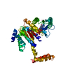
| ||||||||
|---|---|---|---|---|---|---|---|---|---|
| 1 | 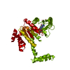
| ||||||||
| Unit cell |
|
-Components
| #1: Protein | Mass: 38596.938 Da / Num. of mol.: 1 Source method: isolated from a genetically manipulated source Source: (gene. exp.) Mycobacterium tuberculosis (bacteria) / Strain: H37Rv / Gene: Rv1496 / Plasmid: AVA0421 / Production host: | ||
|---|---|---|---|
| #2: Chemical | ChemComp-GDP /   Type: RNA linking / Mass: 443.201 Da / Num. of mol.: 1 / Source method: obtained synthetically / Formula: C10H15N5O11P2 / Comment: GDP, energy-carrying molecule*YM Type: RNA linking / Mass: 443.201 Da / Num. of mol.: 1 / Source method: obtained synthetically / Formula: C10H15N5O11P2 / Comment: GDP, energy-carrying molecule*YM | ||
| #3: Chemical | ChemComp-UNX /   Num. of mol.: 4 / Source method: obtained synthetically Num. of mol.: 4 / Source method: obtained synthetically#4: Water | ChemComp-HOH / |  Mass: 18.015 Da / Num. of mol.: 80 / Source method: isolated from a natural source / Formula: H2O Mass: 18.015 Da / Num. of mol.: 80 / Source method: isolated from a natural source / Formula: H2O |
-Experimental details
-Experiment
| Experiment | Method: X-RAY DIFFRACTION / Number of used crystals: 1 |
|---|
- Sample preparation
| Crystal | Density Matthews: 2.63 Å3/Da / Density % sol: 53.27 % |
|---|---|
| Crystal grow | Temperature: 289 K / Method: vapor diffusion, sitting drop / pH: 6.2 Details: 50% PEG 200, 0.2 M NaCl, 0.1 M Na K phosphate pH 6.2, 22.0 mg/mL protein, 2 mM GDP, crystal tracking ID 207391d3, VAPOR DIFFUSION, SITTING DROP, temperature 289K |
-Data collection
| Diffraction | Mean temperature: 100 K |
|---|---|
| Diffraction source | Source: ROTATING ANODE / Type: RIGAKU MICROMAX-007 HF / Wavelength: 1.5418 Å |
| Detector | Type: RIGAKU SATURN 944 / Detector: CCD / Date: Mar 24, 2010 |
| Radiation | Protocol: SINGLE WAVELENGTH / Monochromatic (M) / Laue (L): M / Scattering type: x-ray |
| Radiation wavelength | Wavelength: 1.5418 Å / Relative weight: 1 |
| Reflection | Resolution: 2.45→19.96 Å / Num. obs: 15339 / % possible obs: 99.4 % / Observed criterion σ(I): -3 / Redundancy: 6.84 % / Biso Wilson estimate: 50.04 Å2 / Rmerge(I) obs: 0.072 / Net I/σ(I): 20.5 |
| Reflection shell | Resolution: 2.45→2.51 Å / Redundancy: 4.62 % / Rmerge(I) obs: 0.573 / Mean I/σ(I) obs: 2.7 / Num. measured obs: 5035 / Num. unique obs: 1090 / % possible all: 97.2 |
-Phasing
| Phasing MR |
|
|---|
- Processing
| Software |
| ||||||||||||||||||||||||||||||||||||||||||||||||||||||||||||||||||||||||||||||||||||||||||||||||||||||||||||||||||||||||||||||||||||||||||||||||||||||
|---|---|---|---|---|---|---|---|---|---|---|---|---|---|---|---|---|---|---|---|---|---|---|---|---|---|---|---|---|---|---|---|---|---|---|---|---|---|---|---|---|---|---|---|---|---|---|---|---|---|---|---|---|---|---|---|---|---|---|---|---|---|---|---|---|---|---|---|---|---|---|---|---|---|---|---|---|---|---|---|---|---|---|---|---|---|---|---|---|---|---|---|---|---|---|---|---|---|---|---|---|---|---|---|---|---|---|---|---|---|---|---|---|---|---|---|---|---|---|---|---|---|---|---|---|---|---|---|---|---|---|---|---|---|---|---|---|---|---|---|---|---|---|---|---|---|---|---|---|---|---|---|
| Refinement | Method to determine structure: MOLECULAR REPLACEMENT Starting model: 2qm7 Resolution: 2.45→19.96 Å / Cor.coef. Fo:Fc: 0.939 / Cor.coef. Fo:Fc free: 0.927 / WRfactor Rfree: 0.223 / WRfactor Rwork: 0.184 / Occupancy max: 1 / Occupancy min: 0.5 / FOM work R set: 0.821 / SU B: 15.932 / SU ML: 0.172 / SU R Cruickshank DPI: 0.354 / SU Rfree: 0.251 / Cross valid method: THROUGHOUT / σ(F): 0 / ESU R: 0.354 / ESU R Free: 0.251 / Stereochemistry target values: MAXIMUM LIKELIHOOD Details: HYDROGENS HAVE BEEN ADDED IN THE RIDING POSITIONS; U VALUES: WITH TLS ADDED
| ||||||||||||||||||||||||||||||||||||||||||||||||||||||||||||||||||||||||||||||||||||||||||||||||||||||||||||||||||||||||||||||||||||||||||||||||||||||
| Solvent computation | Ion probe radii: 0.8 Å / Shrinkage radii: 0.8 Å / VDW probe radii: 1.4 Å / Solvent model: MASK | ||||||||||||||||||||||||||||||||||||||||||||||||||||||||||||||||||||||||||||||||||||||||||||||||||||||||||||||||||||||||||||||||||||||||||||||||||||||
| Displacement parameters | Biso max: 121.64 Å2 / Biso mean: 45.656 Å2 / Biso min: 15.58 Å2
| ||||||||||||||||||||||||||||||||||||||||||||||||||||||||||||||||||||||||||||||||||||||||||||||||||||||||||||||||||||||||||||||||||||||||||||||||||||||
| Refinement step | Cycle: LAST / Resolution: 2.45→19.96 Å
| ||||||||||||||||||||||||||||||||||||||||||||||||||||||||||||||||||||||||||||||||||||||||||||||||||||||||||||||||||||||||||||||||||||||||||||||||||||||
| Refine LS restraints |
| ||||||||||||||||||||||||||||||||||||||||||||||||||||||||||||||||||||||||||||||||||||||||||||||||||||||||||||||||||||||||||||||||||||||||||||||||||||||
| LS refinement shell | Resolution: 2.45→2.514 Å / Total num. of bins used: 20
| ||||||||||||||||||||||||||||||||||||||||||||||||||||||||||||||||||||||||||||||||||||||||||||||||||||||||||||||||||||||||||||||||||||||||||||||||||||||
| Refinement TLS params. | Method: refined / Refine-ID: X-RAY DIFFRACTION
| ||||||||||||||||||||||||||||||||||||||||||||||||||||||||||||||||||||||||||||||||||||||||||||||||||||||||||||||||||||||||||||||||||||||||||||||||||||||
| Refinement TLS group |
|
