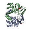
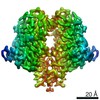
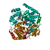
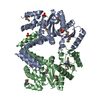
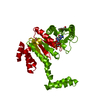
Entry Database : PDB / ID : 4jycTitle MeaB, A Bacterial Homolog of MMAA, in its Apo form Methylmalonyl-CoA mutase accessory protein Keywords / / / / / Function / homology Function Domain/homology Component
/ / / / / / / / / / / / / / / / / / / / / / Biological species Methylobacterium extorquens (bacteria)Method / / / Resolution : 2.2 Å Authors Koutmos, M. / Lofgren, M. / Padovani, D. / Banerjee, R. Journal : Nat.Chem.Biol. / Year : 2013Title : A switch III motif relays signaling between a B12 enzyme and its G-protein chaperone.Authors : Lofgren, M. / Padovani, D. / Koutmos, M. / Banerjee, R. History Deposition Mar 29, 2013 Deposition site / Processing site Revision 1.0 Jul 24, 2013 Provider / Type Revision 1.1 Aug 14, 2013 Group Revision 1.2 Sep 11, 2013 Group Revision 1.3 Sep 20, 2023 Group Data collection / Database references ... Data collection / Database references / Derived calculations / Refinement description Category chem_comp_atom / chem_comp_bond ... chem_comp_atom / chem_comp_bond / database_2 / pdbx_initial_refinement_model / struct_ref_seq_dif / struct_site Item _database_2.pdbx_DOI / _database_2.pdbx_database_accession ... _database_2.pdbx_DOI / _database_2.pdbx_database_accession / _struct_ref_seq_dif.details / _struct_site.pdbx_auth_asym_id / _struct_site.pdbx_auth_comp_id / _struct_site.pdbx_auth_seq_id
Show all Show less
 Movie
Movie Controller


 Open data
 Basic information
 Components
 Keywords
 Function and homology information
 Methylobacterium extorquens (bacteria)
 X-RAY DIFFRACTION /
  Authors
 Citation
 Structure visualization
 Downloads & links
 Other downloads


 PDBj
PDBj
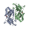
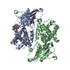
 Assembly
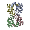


 Type: RNA linking / Mass: 443.201 Da / Num. of mol.: 1 / Source method: obtained synthetically / Formula: C10H15N5O11P2 / Comment: GDP, energy-carrying molecule*YM
 Mass: 18.015 Da / Num. of mol.: 403 / Source method: isolated from a natural source / Formula: H2O
 Sample preparation
 / Beamline: 23-ID-D / Wavelength: 1.03262 Å
 Processing