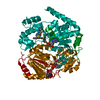
[English] 日本語
Yorodumi
- PDB-5dy4: Crystal structure of human Sirt2 in complex with a brominated 2nd... -

+
Open data


ID or keywords:

Loading...

-
Basic information

Entry
Database: PDB / ID: 5dy4
TitleCrystal structure of human Sirt2 in complex with a brominated 2nd generation SirReal inhibitor and NAD+
ComponentsNAD-dependent protein deacetylase sirtuin-2
KeywordsHYDROLASE / hydrolase inhibitor complex
Function / homology
Function and homology information


cellular response to caloric restriction / negative regulation of oligodendrocyte progenitor proliferation / cellular lipid catabolic process / negative regulation of striated muscle tissue development / NAD-dependent histone H4K16 deacetylase activity / positive regulation of meiotic nuclear division / positive regulation of attachment of spindle microtubules to kinetochore / NAD-dependent protein demyristoylase activity / NAD-dependent protein depalmitoylase activity / paranodal junction ...cellular response to caloric restriction / negative regulation of oligodendrocyte progenitor proliferation / cellular lipid catabolic process / negative regulation of striated muscle tissue development / NAD-dependent histone H4K16 deacetylase activity / positive regulation of meiotic nuclear division / positive regulation of attachment of spindle microtubules to kinetochore / NAD-dependent protein demyristoylase activity / NAD-dependent protein depalmitoylase activity / paranodal junction / tubulin deacetylation / lateral loop / NLRP3 inflammasome complex assembly / peptidyl-lysine deacetylation / mitotic nuclear membrane reassembly / negative regulation of NLRP3 inflammasome complex assembly / tubulin deacetylase activity / regulation of exit from mitosis / paranode region of axon / NAD-dependent protein lysine deacetylase activity / Schmidt-Lanterman incisure / positive regulation of fatty acid biosynthetic process / myelination in peripheral nervous system / rDNA heterochromatin formation / protein acetyllysine N-acetyltransferase / chromatin silencing complex / regulation of phosphorylation / Initiation of Nuclear Envelope (NE) Reformation / protein deacetylation / NAD-dependent histone deacetylase activity / positive regulation of oocyte maturation / juxtaparanode region of axon / protein lysine deacetylase activity / meiotic spindle / response to redox state / regulation of myelination / histone acetyltransferase binding / histone deacetylase activity / negative regulation of fat cell differentiation / negative regulation of peptidyl-threonine phosphorylation / positive regulation of execution phase of apoptosis / glial cell projection / positive regulation of cell division / NAD+-protein poly-ADP-ribosyltransferase activity / NAD+ binding / subtelomeric heterochromatin formation / negative regulation of reactive oxygen species metabolic process / positive regulation of DNA binding / heterochromatin / heterochromatin formation / epigenetic regulation of gene expression / Transferases; Acyltransferases; Transferring groups other than aminoacyl groups / cellular response to epinephrine stimulus / substantia nigra development / centriole / negative regulation of autophagy / meiotic cell cycle / ubiquitin binding / negative regulation of protein catabolic process / mitotic spindle / autophagy / histone deacetylase binding / spindle / positive regulation of proteasomal ubiquitin-dependent protein catabolic process / myelin sheath / chromosome / midbody / cellular response to oxidative stress / growth cone / cellular response to hypoxia / perikaryon / proteasome-mediated ubiquitin-dependent protein catabolic process / DNA-binding transcription factor binding / microtubule / chromosome, telomeric region / regulation of cell cycle / cell division / innate immune response / negative regulation of DNA-templated transcription / centrosome / chromatin binding / nucleolus / perinuclear region of cytoplasm / negative regulation of transcription by RNA polymerase II / positive regulation of transcription by RNA polymerase II / mitochondrion / zinc ion binding / nucleus / plasma membrane / cytoplasm / cytosol
Similarity search - Function
Sirtuin, class I / SIR2/SIRT2 'Small Domain' / SIR2/SIRT2 'Small Domain' / Sirtuin, catalytic core small domain superfamily / Sirtuin family / Sir2 family / Sirtuin family, catalytic core domain / Sirtuin catalytic domain profile. / TPP-binding domain / DHS-like NAD/FAD-binding domain superfamily ...Sirtuin, class I / SIR2/SIRT2 'Small Domain' / SIR2/SIRT2 'Small Domain' / Sirtuin, catalytic core small domain superfamily / Sirtuin family / Sir2 family / Sirtuin family, catalytic core domain / Sirtuin catalytic domain profile. / TPP-binding domain / DHS-like NAD/FAD-binding domain superfamily / Rossmann fold / 2-Layer Sandwich / 3-Layer(aba) Sandwich / Alpha Beta
Similarity search - Domain/homology
Chem-5GN / NICOTINAMIDE-ADENINE-DINUCLEOTIDE / NAD-dependent protein deacetylase sirtuin-2
Similarity search - Component
Biological speciesHomo sapiens (human)
MethodX-RAY DIFFRACTION / SYNCHROTRON / MOLECULAR REPLACEMENT / Resolution: 1.77 Å
AuthorsRumpf, T. / Gerhardt, S. / Einsle, O. / Jung, M.
Funding support Germany, 1items
OrganizationGrant numberCountry
German Research FoundationJu295/8-1 Germany
CitationJournal: J.Med.Chem. / Year: 2016
Title: Aminothiazoles as Potent and Selective Sirt2 Inhibitors: A Structure-Activity Relationship Study.
Authors: Schiedel, M. / Rumpf, T. / Karaman, B. / Lehotzky, A. / Olah, J. / Gerhardt, S. / Ovadi, J. / Sippl, W. / Einsle, O. / Jung, M.
History
DepositionSep 24, 2015Deposition site: RCSB / Processing site: PDBE
Revision 1.0Jan 13, 2016Provider: repository / Type: Initial release
Revision 1.1Jan 20, 2016Group: Database references
Revision 1.2Mar 9, 2016Group: Database references
Revision 1.3Jan 10, 2024Group: Author supporting evidence / Data collection ...Author supporting evidence / Data collection / Database references / Refinement description
Category: chem_comp_atom / chem_comp_bond ...chem_comp_atom / chem_comp_bond / database_2 / pdbx_audit_support / pdbx_initial_refinement_model
Item: _database_2.pdbx_DOI / _database_2.pdbx_database_accession / _pdbx_audit_support.funding_organization

-
Structure visualization

Structure viewerMolecule:
MolmilJmol/JSmol

Downloads & links

-
Assembly

Deposited unit
A: NAD-dependent protein deacetylase sirtuin-2
hetero molecules


Theoretical massNumber of molelcules
Total (without water)35,6454
Polymers34,4171
Non-polymers1,2283
Water3,297183
1
A: NAD-dependent protein deacetylase sirtuin-2
hetero molecules

A: NAD-dependent protein deacetylase sirtuin-2
hetero molecules


Theoretical massNumber of molelcules
Total (without water)71,2908
Polymers68,8332
Non-polymers2,4576
Water362
TypeNameSymmetry operationNumber
identity operation1_555x,y,z1
crystal symmetry operation2_555-x,y,-z1
Buried area5880 Å2
ΔGint-43 kcal/mol
Surface area25520 Å2
MethodPISA
2


  • Idetical with deposited unit
  • defined by author&software
TypeNameSymmetry operationNumber
identity operation1_555x,y,z1
Buried area1200 Å2
ΔGint-3 kcal/mol
Surface area14490 Å2
MethodPISA
Unit cell
Length a, b, c (Å)84.303, 55.432, 96.176
Angle α, β, γ (deg.)90.00, 114.91, 90.00
Int Tables number5
Space group name H-MI121
Components on special symmetry positions
IDModelComponents
11A-663-

HOH

21A-683-

HOH

-
Components

#1: Protein NAD-dependent protein deacetylase sirtuin-2 / Regulatory protein SIR2 homolog 2 / SIR2-like protein 2


Mass: 34416.727 Da / Num. of mol.: 1 / Fragment: residues 56-356
Source method: isolated from a genetically manipulated source
Source: (gene. exp.) Homo sapiens (human) / Gene: SIRT2, SIR2L, SIR2L2 / Plasmid: modified pET15b / Production host: Escherichia coli BL21(DE3) (bacteria) / Variant (production host): Codonplus RIPL
References: UniProt: Q8IXJ6, Hydrolases; Acting on carbon-nitrogen bonds, other than peptide bonds; In linear amides
#2: Chemical ChemComp-ZN / ZINC ION


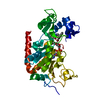
Mass: 65.409 Da / Num. of mol.: 1 / Source method: obtained synthetically / Formula: Zn
#3: Chemical ChemComp-5GN / N-{5-[(7-bromonaphthalen-1-yl)methyl]-1,3-thiazol-2-yl}-2-[(4,6-dimethylpyrimidin-2-yl)sulfanyl]acetamide


Mass: 499.446 Da / Num. of mol.: 1 / Source method: obtained synthetically / Formula: C22H19BrN4OS2
#4: Chemical ChemComp-NAD / NICOTINAMIDE-ADENINE-DINUCLEOTIDE


Mass: 663.425 Da / Num. of mol.: 1 / Source method: obtained synthetically / Formula: C21H27N7O14P2 / Comment: NAD*YM
#5: Water ChemComp-HOH / water


Mass: 18.015 Da / Num. of mol.: 183 / Source method: isolated from a natural source / Formula: H2O

-
Experimental details

-
Experiment

ExperimentMethod: X-RAY DIFFRACTION

-
Sample preparation

CrystalDensity Matthews: 2.96 Å3/Da / Density % sol: 58.46 %
Crystal growTemperature: 278 K / Method: vapor diffusion, sitting drop / pH: 7 / Details: 27 % PEG 3350 / PH range: 7

-
Data collection

DiffractionMean temperature: 100 K
Diffraction sourceSource: SYNCHROTRON / Site: SLS / Beamline: X06SA / Wavelength: 1 Å
DetectorType: DECTRIS PILATUS 6M / Detector: PIXEL / Date: Oct 30, 2014
RadiationProtocol: SINGLE WAVELENGTH / Monochromatic (M) / Laue (L): M / Scattering type: x-ray
Radiation wavelengthWavelength: 1 Å / Relative weight: 1
ReflectionResolution: 1.77→46.78 Å / Num. obs: 38622 / % possible obs: 98.2 % / Redundancy: 6.7 % / Rmerge(I) obs: 0.095 / Net I/σ(I): 10.3
Reflection shellResolution: 1.77→1.81 Å / Redundancy: 6.9 % / Rmerge(I) obs: 2.05 / Mean I/σ(I) obs: 1 / % possible all: 98.1

-
Processing

Software
NameVersionClassification
REFMAC5.8.0131refinement
XDSdata reduction
Aimlessdata scaling
MOLREPphasing
RefinementMethod to determine structure: MOLECULAR REPLACEMENT
Starting model: 4RMG
Resolution: 1.77→46.78 Å / Cor.coef. Fo:Fc: 0.973 / Cor.coef. Fo:Fc free: 0.95 / SU B: 5.857 / SU ML: 0.094 / Cross valid method: THROUGHOUT / ESU R: 0.101 / ESU R Free: 0.104 / Stereochemistry target values: MAXIMUM LIKELIHOOD / Details: HYDROGENS HAVE BEEN ADDED IN THE RIDING POSITIONS
RfactorNum. reflection% reflectionSelection details
Rfree0.21286 1913 5 %RANDOM
Rwork0.17494 ---
obs0.17686 36691 97.9 %-
Solvent computationIon probe radii: 0.8 Å / Shrinkage radii: 0.8 Å / VDW probe radii: 1.2 Å / Solvent model: MASK
Displacement parametersBiso mean: 33.455 Å2
Baniso -1Baniso -2Baniso -3
1--1.09 Å20 Å2-1 Å2
2--1.34 Å20 Å2
3---0.47 Å2
Refinement stepCycle: 1 / Resolution: 1.77→46.78 Å
ProteinNucleic acidLigandSolventTotal
Num. atoms2391 0 75 183 2649
Refine LS restraints
Refine-IDTypeDev idealDev ideal targetNumber
X-RAY DIFFRACTIONr_bond_refined_d0.0130.0192531
X-RAY DIFFRACTIONr_bond_other_d0.0020.022377
X-RAY DIFFRACTIONr_angle_refined_deg1.6461.9943420
X-RAY DIFFRACTIONr_angle_other_deg1.0213.0015489
X-RAY DIFFRACTIONr_dihedral_angle_1_deg6.1555299
X-RAY DIFFRACTIONr_dihedral_angle_2_deg34.24923.67109
X-RAY DIFFRACTIONr_dihedral_angle_3_deg12.63315438
X-RAY DIFFRACTIONr_dihedral_angle_4_deg16.2051515
X-RAY DIFFRACTIONr_chiral_restr0.0980.2364
X-RAY DIFFRACTIONr_gen_planes_refined0.0070.0212906
X-RAY DIFFRACTIONr_gen_planes_other0.0020.02574
X-RAY DIFFRACTIONr_nbd_refined
X-RAY DIFFRACTIONr_nbd_other
X-RAY DIFFRACTIONr_nbtor_refined
X-RAY DIFFRACTIONr_nbtor_other
X-RAY DIFFRACTIONr_xyhbond_nbd_refined
X-RAY DIFFRACTIONr_xyhbond_nbd_other
X-RAY DIFFRACTIONr_metal_ion_refined
X-RAY DIFFRACTIONr_metal_ion_other
X-RAY DIFFRACTIONr_symmetry_vdw_refined
X-RAY DIFFRACTIONr_symmetry_vdw_other
X-RAY DIFFRACTIONr_symmetry_hbond_refined
X-RAY DIFFRACTIONr_symmetry_hbond_other
X-RAY DIFFRACTIONr_symmetry_metal_ion_refined
X-RAY DIFFRACTIONr_symmetry_metal_ion_other
X-RAY DIFFRACTIONr_mcbond_it1.4842.7361204
X-RAY DIFFRACTIONr_mcbond_other1.4842.7361203
X-RAY DIFFRACTIONr_mcangle_it2.4214.0871500
X-RAY DIFFRACTIONr_mcangle_other2.4214.0871501
X-RAY DIFFRACTIONr_scbond_it1.8973.061327
X-RAY DIFFRACTIONr_scbond_other1.8963.061328
X-RAY DIFFRACTIONr_scangle_it
X-RAY DIFFRACTIONr_scangle_other3.1014.4821921
X-RAY DIFFRACTIONr_long_range_B_refined5.08822.9312946
X-RAY DIFFRACTIONr_long_range_B_other5.02722.5772872
X-RAY DIFFRACTIONr_rigid_bond_restr
X-RAY DIFFRACTIONr_sphericity_free
X-RAY DIFFRACTIONr_sphericity_bonded
LS refinement shellResolution: 1.77→1.816 Å / Total num. of bins used: 20
RfactorNum. reflection% reflection
Rfree0.335 142 -
Rwork0.306 2726 -
obs--97.98 %
Refinement TLS params.Method: refined / Origin x: -15.9435 Å / Origin y: -26.8785 Å / Origin z: -0.054 Å
111213212223313233
T0.012 Å20.0244 Å20.009 Å2-0.0602 Å20.0255 Å2--0.0199 Å2
L0.4732 °20.0327 °20.278 °2-0.0523 °2-0.0799 °2--0.3732 °2
S0.0147 Å °0.0146 Å °0.0122 Å °0.0019 Å °0.0237 Å °0.0269 Å °0.0116 Å °-0.0097 Å °-0.0385 Å °

+
About Yorodumi

-
News

-
Feb 9, 2022. New format data for meta-information of EMDB entries

New format data for meta-information of EMDB entries

  • Version 3 of the EMDB header file is now the official format.
  • The previous official version 1.9 will be removed from the archive.

Related info.:EMDB header

External links:wwPDB to switch to version 3 of the EMDB data model

-
Aug 12, 2020. Covid-19 info

Covid-19 info

URL: https://pdbjlvh1.pdbj.org/emnavi/covid19.php

New page: Covid-19 featured information page in EM Navigator.

Related info.:Covid-19 info / Mar 5, 2020. Novel coronavirus structure data

+
Mar 5, 2020. Novel coronavirus structure data

Novel coronavirus structure data

Related info.:Yorodumi Speices / Aug 12, 2020. Covid-19 info

External links:COVID-19 featured content - PDBj / Molecule of the Month (242):Coronavirus Proteases

+
Jan 31, 2019. EMDB accession codes are about to change! (news from PDBe EMDB page)

EMDB accession codes are about to change! (news from PDBe EMDB page)

  • The allocation of 4 digits for EMDB accession codes will soon come to an end. Whilst these codes will remain in use, new EMDB accession codes will include an additional digit and will expand incrementally as the available range of codes is exhausted. The current 4-digit format prefixed with “EMD-” (i.e. EMD-XXXX) will advance to a 5-digit format (i.e. EMD-XXXXX), and so on. It is currently estimated that the 4-digit codes will be depleted around Spring 2019, at which point the 5-digit format will come into force.
  • The EM Navigator/Yorodumi systems omit the EMD- prefix.

Related info.:Q: What is EMD? / ID/Accession-code notation in Yorodumi/EM Navigator

External links:EMDB Accession Codes are Changing Soon! / Contact to PDBj

+
Jul 12, 2017. Major update of PDB

Major update of PDB

  • wwPDB released updated PDB data conforming to the new PDBx/mmCIF dictionary.
  • This is a major update changing the version number from 4 to 5, and with Remediation, in which all the entries are updated.
  • In this update, many items about electron microscopy experimental information are reorganized (e.g. em_software).
  • Now, EM Navigator and Yorodumi are based on the updated data.

External links:wwPDB Remediation / Enriched Model Files Conforming to OneDep Data Standards Now Available in the PDB FTP Archive

-
Yorodumi

Thousand views of thousand structures

  • Yorodumi is a browser for structure data from EMDB, PDB, SASBDB, etc.
  • This page is also the successor to EM Navigator detail page, and also detail information page/front-end page for Omokage search.
  • The word "yorodu" (or yorozu) is an old Japanese word meaning "ten thousand". "mi" (miru) is to see.

Related info.:EMDB / PDB / SASBDB / Comparison of 3 databanks / Yorodumi Search / Aug 31, 2016. New EM Navigator & Yorodumi / Yorodumi Papers / Jmol/JSmol / Function and homology information / Changes in new EM Navigator and Yorodumi

Read more