 Movie
Movie Controller

[English] 日本語
 Yorodumi
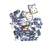
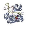
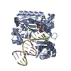
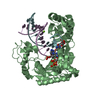
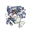
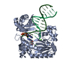
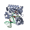
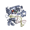
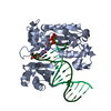
- PDB-3m9m: Crystal Structure of Dpo4 in complex with DNA containing the majo... -
+ Open data

- Basic information
| Entry | Database: PDB / ID: 3m9m | ||||||
|---|---|---|---|---|---|---|---|
| Title | Crystal Structure of Dpo4 in complex with DNA containing the major cisplatin lesion | ||||||
 Components Components |
| ||||||
 Keywords Keywords | Transferase/DNA / Dpo4 / translesion DNA synthesis / TLS / cisplatin / Y-family DNA polymerase / protein-DNA complex / DNA / DNA damage / DNA repair / DNA replication / DNA-binding / DNA-directed DNA polymerase / Magnesium / Metal-binding / Mutator protein / Nucleotidyltransferase / Transferase / Transferase-DNA complex | ||||||
| Function / homology |  Function and homology information Function and homology informationerror-prone translesion synthesis / DNA-templated DNA replication / DNA-directed DNA polymerase / damaged DNA binding / DNA-directed DNA polymerase activity / DNA repair / magnesium ion binding / cytoplasm Similarity search - Function | ||||||
| Biological species |   Sulfolobus solfataricus (archaea) Sulfolobus solfataricus (archaea)Synthetic construct (others) | ||||||
| Method |  X-RAY DIFFRACTION / SYNCHROTRON / MOLECULAR REPLACEMENT / Resolution: 2.9 Å X-RAY DIFFRACTION / SYNCHROTRON / MOLECULAR REPLACEMENT / Resolution: 2.9 Å | ||||||
 Authors Authors | Wong, J.H.Y. / Ling, H. | ||||||
 Citation Citation | Journal: Embo J. / Year: 2010 Title: Structural insight into dynamic bypass of the major cisplatin-DNA adduct by Y-family polymerase Dpo4. Authors: Wong, J.H. / Brown, J.A. / Suo, Z. / Blum, P. / Nohmi, T. / Ling, H. | ||||||
| History |
|
- Structure visualization
| Structure viewer | Molecule: MolmilJmol/JSmol |
|---|
- Downloads & links
-Download
| PDBx/mmCIF format | 3m9m.cif.gz | 103.4 KB | Display | PDBx/mmCIF format |
|---|---|---|---|---|
| PDB format | pdb3m9m.ent.gz | 74.8 KB | Display | PDB format |
| PDBx/mmJSON format | 3m9m.json.gz | Tree view | PDBx/mmJSON format | |
| Others |  Other downloads Other downloads |
-Validation report
| Arichive directory | https://data.pdbj.org/pub/pdb/validation_reports/m9/3m9mftp://data.pdbj.org/pub/pdb/validation_reports/m9/3m9m | HTTPS FTP |
|---|
-Related structure data
| Related structure data |  3m9nC  3m9oC 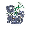 1jx4S C: citing same article ( S: Starting model for refinement |
|---|---|
| Similar structure data |


-Links
 PDBj
PDBj


- Assembly
| Deposited unit | 
| ||||||||
|---|---|---|---|---|---|---|---|---|---|
| 1 |
| ||||||||
| Unit cell |
|
-Components
-Protein , 1 types, 1 molecules B
| #1: Protein | Mass: 40257.879 Da / Num. of mol.: 1 Source method: isolated from a genetically manipulated source Source: (gene. exp.) Sulfolobus solfataricus (archaea) / Strain: P2 / Gene: dbh, dpo4, SSO2448 / Plasmid: pET22b / Production host:  |
|---|
-DNA chain , 2 types, 2 molecules PT
| #2: DNA chain | Mass: 4154.722 Da / Num. of mol.: 1 / Source method: obtained synthetically / Source: (synth.) Synthetic construct (others) |
|---|---|
| #3: DNA chain | Mass: 5345.439 Da / Num. of mol.: 1 / Source method: obtained synthetically / Source: (synth.) Synthetic construct (others) |
-Non-polymers , 5 types, 60 molecules 


| #4: Chemical | ChemComp-CTP /  Mass: 483.156 Da / Num. of mol.: 1 / Source method: obtained synthetically / Formula: C9H16N3O14P3 Mass: 483.156 Da / Num. of mol.: 1 / Source method: obtained synthetically / Formula: C9H16N3O14P3 | ||||||
|---|---|---|---|---|---|---|---|
| #5: Chemical | ChemComp-CA /  Mass: 40.078 Da / Num. of mol.: 4 / Source method: obtained synthetically / Formula: Ca Mass: 40.078 Da / Num. of mol.: 4 / Source method: obtained synthetically / Formula: Ca#6: Chemical |  Mass: 92.094 Da / Num. of mol.: 3 / Source method: obtained synthetically / Formula: C3H8O3 Mass: 92.094 Da / Num. of mol.: 3 / Source method: obtained synthetically / Formula: C3H8O3#7: Chemical | ChemComp-CPT / |  Mass: 300.045 Da / Num. of mol.: 1 / Source method: obtained synthetically / Formula: Cl2H6N2Pt / Comment: medication*YM Mass: 300.045 Da / Num. of mol.: 1 / Source method: obtained synthetically / Formula: Cl2H6N2Pt / Comment: medication*YM#8: Water | ChemComp-HOH / | Mass: 18.015 Da / Num. of mol.: 51 / Source method: isolated from a natural source / Formula: H2O |
-Experimental details
-Experiment
| Experiment | Method: X-RAY DIFFRACTION / Number of used crystals: 1 |
|---|
- Sample preparation
| Crystal | Density Matthews: 2.63 Å3/Da / Density % sol: 53.18 % |
|---|---|
| Crystal grow | Temperature: 298 K / Method: hanging drop / pH: 7.5 Details: 0.2M calcium acetate, 5% glycerol, 15% PEG 3350, 200mM NaCl, pH 7.5, hanging drop, temperature 298K |
-Data collection
| Diffraction | Mean temperature: 100 K | ||||||||||||||||||||||||||||||||||||||||||||||||||||||||||||||||||
|---|---|---|---|---|---|---|---|---|---|---|---|---|---|---|---|---|---|---|---|---|---|---|---|---|---|---|---|---|---|---|---|---|---|---|---|---|---|---|---|---|---|---|---|---|---|---|---|---|---|---|---|---|---|---|---|---|---|---|---|---|---|---|---|---|---|---|---|
| Diffraction source | Source: SYNCHROTRON / Site: APS  / Beamline: 24-ID-C / Wavelength: 1.0781 Å / Beamline: 24-ID-C / Wavelength: 1.0781 Å | ||||||||||||||||||||||||||||||||||||||||||||||||||||||||||||||||||
| Detector | Type: ADSC Q315 / Detector: CCD / Date: Mar 12, 2008 | ||||||||||||||||||||||||||||||||||||||||||||||||||||||||||||||||||
| Radiation | Monochromator: Cryo-Cooled Si(111) double crystal / Protocol: SINGLE WAVELENGTH / Monochromatic (M) / Laue (L): M / Scattering type: x-ray | ||||||||||||||||||||||||||||||||||||||||||||||||||||||||||||||||||
| Radiation wavelength | Wavelength: 1.0781 Å / Relative weight: 1 | ||||||||||||||||||||||||||||||||||||||||||||||||||||||||||||||||||
| Reflection | Resolution: 2.9→50 Å / Num. all: 12180 / Num. obs: 12009 / % possible obs: 98.6 % / Observed criterion σ(F): 1 / Observed criterion σ(I): 1 / Redundancy: 3.5 % / Rmerge(I) obs: 0.061 / Χ2: 1.161 / Net I/σ(I): 10.7 | ||||||||||||||||||||||||||||||||||||||||||||||||||||||||||||||||||
| Reflection shell |
|
- Processing
| Software |
| ||||||||||||||||||||||||||||
|---|---|---|---|---|---|---|---|---|---|---|---|---|---|---|---|---|---|---|---|---|---|---|---|---|---|---|---|---|---|
| Refinement | Method to determine structure: MOLECULAR REPLACEMENT Starting model: PDB ENTRY 1JX4 Resolution: 2.9→45 Å / Occupancy max: 1 / Occupancy min: 1 / FOM work R set: 0.748 / Cross valid method: THROUGHOUT / σ(F): 0 / Stereochemistry target values: Engh & Huber
| ||||||||||||||||||||||||||||
| Solvent computation | Bsol: 32.47 Å2 | ||||||||||||||||||||||||||||
| Displacement parameters | Biso max: 137.45 Å2 / Biso mean: 63.213 Å2 / Biso min: 17.73 Å2
| ||||||||||||||||||||||||||||
| Refinement step | Cycle: LAST / Resolution: 2.9→45 Å
| ||||||||||||||||||||||||||||
| Refine LS restraints |
| ||||||||||||||||||||||||||||
| Xplor file |
|
