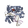
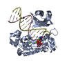
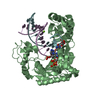
error-prone translesion synthesis / DNA-templated DNA replication / DNA-directed DNA polymerase / damaged DNA binding / DNA-directed DNA polymerase activity / DNA repair / magnesium ion binding / cytoplasm Similarity search - Function
DNA polymerase type-Y, HhH motif / IMS family HHH motif / DNA polymerase IV / : / DNA polymerase, Y-family, little finger domain / MutS, DNA mismatch repair protein, domain I - #60 / MutS, DNA mismatch repair protein, domain I / DNA polymerase, Y-family, little finger domain / impB/mucB/samB family C-terminal domain / UmuC domain ...DNA polymerase type-Y, HhH motif / IMS family HHH motif / DNA polymerase IV / : / DNA polymerase, Y-family, little finger domain / MutS, DNA mismatch repair protein, domain I - #60 / MutS, DNA mismatch repair protein, domain I / DNA polymerase, Y-family, little finger domain / impB/mucB/samB family C-terminal domain / UmuC domain / DNA polymerase, Y-family, little finger domain superfamily / impB/mucB/samB family / UmuC domain profile. / Reverse transcriptase/Diguanylate cyclase domain / Dna Ligase; domain 1 / 5' to 3' exonuclease, C-terminal subdomain / DNA polymerase; domain 1 / Reverse transcriptase/Diguanylate cyclase domain / Alpha-Beta Plaits / DNA/RNA polymerase superfamily / 2-Layer Sandwich / Orthogonal Bundle / 3-Layer(aba) Sandwich / Mainly Alpha / Alpha Beta Similarity search - Domain/homology
Resolution: 1.7→1.73 Å / Redundancy: 4.34 % / Rmerge(I) obs: 0.55 / Mean I/σ(I) obs: 2.2 / Num. unique all: 2815 / Rsym value: 0.554 / % possible all: 98.4
Reflection
*PLUS
Rmerge(I) obs: 0.04
Reflection shell
*PLUS
% possible obs: 98.4 % / Rmerge(I) obs: 0.554
-
Processing
Software
Name
Version
Classification
SOLVE
phasing
CNS
1
refinement
DENZO
datareduction
SCALEPACK
datascaling
Refinement
Method to determine structure: MAD / Resolution: 1.7→500 Å / Isotropic thermal model: Isotropic / Cross valid method: THROUGHOUT / σ(F): 0 / Stereochemistry target values: Engh & Huber Details: mlhl: maximum likelihood target using amplitudes and phase probability distribution
Rfactor
Num. reflection
% reflection
Selection details
Rfree
0.228
1122
-
random
Rwork
0.213
-
-
-
all
0.213
58391
-
-
obs
0.213
57010
97.6 %
-
Displacement parameters
Biso mean: 34.9 Å2
Baniso -1
Baniso -2
Baniso -3
1-
-9.07 Å2
0 Å2
0 Å2
2-
-
2.618 Å2
0 Å2
3-
-
-
6.458 Å2
Refinement step
Cycle: LAST / Resolution: 1.7→500 Å
Protein
Nucleic acid
Ligand
Solvent
Total
Num. atoms
2791
611
27
525
3954
Refine LS restraints
Refine-ID
Type
Dev ideal
X-RAY DIFFRACTION
c_bond_d
0.017
X-RAY DIFFRACTION
c_angle_deg
1.943
LS refinement shell
Resolution: 1.7→1.73 Å / Rfactor Rfree error: 0.007
In the structure databanks used in Yorodumi, some data are registered as the other names, "COVID-19 virus" and "2019-nCoV". Here are the details of the virus and the list of structure data.
Jan 31, 2019. EMDB accession codes are about to change! (news from PDBe EMDB page)
EMDB accession codes are about to change! (news from PDBe EMDB page)
The allocation of 4 digits for EMDB accession codes will soon come to an end. Whilst these codes will remain in use, new EMDB accession codes will include an additional digit and will expand incrementally as the available range of codes is exhausted. The current 4-digit format prefixed with “EMD-” (i.e. EMD-XXXX) will advance to a 5-digit format (i.e. EMD-XXXXX), and so on. It is currently estimated that the 4-digit codes will be depleted around Spring 2019, at which point the 5-digit format will come into force.
The EM Navigator/Yorodumi systems omit the EMD- prefix.
Related info.:Q: What is EMD? / ID/Accession-code notation in Yorodumi/EM Navigator
Yorodumi is a browser for structure data from EMDB, PDB, SASBDB, etc.
This page is also the successor to EM Navigator detail page, and also detail information page/front-end page for Omokage search.
The word "yorodu" (or yorozu) is an old Japanese word meaning "ten thousand". "mi" (miru) is to see.
Related info.:EMDB / PDB / SASBDB / Comparison of 3 databanks / Yorodumi Search / Aug 31, 2016. New EM Navigator & Yorodumi / Yorodumi Papers / Jmol/JSmol / Function and homology information / Changes in new EM Navigator and Yorodumi
 Movie
Movie Controller

 Yorodumi
 Open data
 Basic information
 Components
 Keywords
 Function and homology information
 Sulfolobus solfataricus (archaea)
 X-RAY DIFFRACTION /
  Authors
 Citation
 Structure visualization
 Downloads & links
 Other downloads
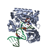


 PDBj
PDBj


 Assembly


 Mass: 40.078 Da / Num. of mol.: 1 / Source method: obtained synthetically / Formula: Ca
 Mass: 24.305 Da / Num. of mol.: 1 / Source method: obtained synthetically / Formula: Mg
 Mass: 395.202 Da / Num. of mol.: 1 / Source method: obtained synthetically / Formula: C10H15N5O8P2
 Sample preparation
 / Beamline: X9B / Wavelength: 0.9832 Å
 Processing