 Movie
Movie Controller

[English] 日本語
 Yorodumi
- PDB-3lg8: Crystal structure of the C-terminal part of subunit E (E101-206) ... -
+ Open data

- Basic information
| Entry | Database: PDB / ID: 3lg8 | ||||||
|---|---|---|---|---|---|---|---|
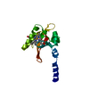
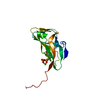
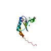
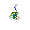
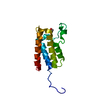
| Title | Crystal structure of the C-terminal part of subunit E (E101-206) from Methanocaldococcus jannaschii of A1AO ATP synthase | ||||||
 Components Components | A-type ATP synthase subunit E | ||||||
 Keywords Keywords | HYDROLASE / Archaea / peripheral stalk / Structural protein / TRANSPORT PROTEIN | ||||||
| Function / homology |  Function and homology information Function and homology informationproton-transporting two-sector ATPase complex, catalytic domain / proton motive force-driven plasma membrane ATP synthesis / proton-transporting ATPase activity, rotational mechanism / proton-transporting ATP synthase activity, rotational mechanism / ATP binding / plasma membrane Similarity search - Function | ||||||
| Biological species |   Methanocaldococcus jannaschii (archaea) Methanocaldococcus jannaschii (archaea) | ||||||
| Method |  X-RAY DIFFRACTION / SYNCHROTRON / MOLECULAR REPLACEMENT / molecular replacement / Resolution: 4.1 Å X-RAY DIFFRACTION / SYNCHROTRON / MOLECULAR REPLACEMENT / molecular replacement / Resolution: 4.1 Å | ||||||
 Authors Authors | Balakrishna, A.M. / Manimekalai, M.S.S. / Hunke, C. / Gayen, S. / Jeyakanthan, J. / Gruber, G. | ||||||
 Citation Citation | Journal: J.Bioenerg.Biomembr. / Year: 2010 Title: Crystal and solution structure of the C-terminal part of the Methanocaldococcus jannaschii A1AO ATP synthase subunit E revealed by X-ray diffraction and small-angle X-ray scattering Authors: Balakrishna, A.M. / Manimekalai, M.S.S. / Hunke, C. / Gayen, S. / Rossle, M. / Jeyakanthan, J. / Gruber, G. #1: Journal: J.Mol.Biol. / Year: 2007 Title: Dimeric core structure of modular stator subunit E of archaeal H+ -ATPase Authors: Lokanath, N.K. / Matsuura, Y. / Kuroishi, C. / Takahashi, N. / Kunishima, N. | ||||||
| History |
|
- Structure visualization
| Structure viewer | Molecule: MolmilJmol/JSmol |
|---|
- Downloads & links
-Download
| PDBx/mmCIF format | 3lg8.cif.gz | 44 KB | Display | PDBx/mmCIF format |
|---|---|---|---|---|
| PDB format | pdb3lg8.ent.gz | 27.6 KB | Display | PDB format |
| PDBx/mmJSON format | 3lg8.json.gz | Tree view | PDBx/mmJSON format | |
| Others |  Other downloads Other downloads |
-Validation report
| Arichive directory | https://data.pdbj.org/pub/pdb/validation_reports/lg/3lg8ftp://data.pdbj.org/pub/pdb/validation_reports/lg/3lg8 | HTTPS FTP |
|---|
-Related structure data
| Related structure data |  2dm9S S: Starting model for refinement |
|---|---|
| Similar structure data |


-Links
 PDBj
PDBj

- Assembly
| Deposited unit | 
| ||||||||||||||||||
|---|---|---|---|---|---|---|---|---|---|---|---|---|---|---|---|---|---|---|---|
| 1 | 
| ||||||||||||||||||
| 2 | 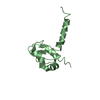
| ||||||||||||||||||
| Unit cell |
| ||||||||||||||||||
| Noncrystallographic symmetry (NCS) | NCS domain:
NCS domain segments: Component-ID: 1 / Ens-ID: 1 / Beg auth comp-ID: TYR / Beg label comp-ID: TYR / End auth comp-ID: ARG / End label comp-ID: ARG / Refine code: 4 / Auth seq-ID: 105 - 200 / Label seq-ID: 5 - 100
|
-Components
| #1: Protein | Mass: 11854.684 Da / Num. of mol.: 2 / Fragment: UNP residues 101-206 Source method: isolated from a genetically manipulated source Source: (gene. exp.) Methanocaldococcus jannaschii (archaea)Strain: ATCC 43067 / Gene: atpE, MJ0220 / Plasmid: pET 9d / Production host:  |
|---|
-Experimental details
-Experiment
| Experiment | Method: X-RAY DIFFRACTION / Number of used crystals: 1 |
|---|
- Sample preparation
| Crystal | Density Matthews: 2.47 Å3/Da / Density % sol: 50.23 % |
|---|---|
| Crystal grow | Temperature: 291 K / Method: vapor diffusion, hanging drop / pH: 6.5 Details: 0.05M Cesium chloride, 0.1M MES monohydrate buffer (pH 6.5), 30% v/v Jeffamine M-600, 1mM TCEP, 0.1mM glycine, VAPOR DIFFUSION, HANGING DROP, temperature 291K |
-Data collection
| Diffraction | Mean temperature: 100 K |
|---|---|
| Diffraction source | Source: SYNCHROTRON / Site: SPring-8  / Beamline: BL12B2 / Wavelength: 1 Å / Beamline: BL12B2 / Wavelength: 1 Å |
| Detector | Type: MARMOSAIC 225 mm CCD / Detector: CCD / Date: Feb 28, 2009 / Details: mirrors |
| Radiation | Monochromator: GRAPHITE / Protocol: SINGLE WAVELENGTH / Monochromatic (M) / Laue (L): M / Scattering type: x-ray |
| Radiation wavelength | Wavelength: 1 Å / Relative weight: 1 |
| Reflection | Resolution: 4.1→30 Å / Num. obs: 2125 / % possible obs: 99.3 % / Observed criterion σ(F): 0 / Observed criterion σ(I): 0 / Redundancy: 10.8 % / Rmerge(I) obs: 0.068 / Net I/σ(I): 27.35 |
| Reflection shell | Resolution: 4.1→4.17 Å / Redundancy: 11.5 % / Rmerge(I) obs: 0.076 / Mean I/σ(I) obs: 26.35 / % possible all: 100 |
-Phasing
| Phasing | Method: molecular replacement |
|---|
- Processing
| Software |
| ||||||||||||||||||||||||||||||
|---|---|---|---|---|---|---|---|---|---|---|---|---|---|---|---|---|---|---|---|---|---|---|---|---|---|---|---|---|---|---|---|
| Refinement | Method to determine structure: MOLECULAR REPLACEMENT Starting model: PDB ENTRY 2DM9 Resolution: 4.1→24.27 Å / Cor.coef. Fo:Fc: 0.803 / Cor.coef. Fo:Fc free: 0.735 / Occupancy max: 1 / Occupancy min: 1 / SU B: 108.693 / SU ML: 1.483 / Isotropic thermal model: overall / Cross valid method: THROUGHOUT / σ(F): 0 / ESU R Free: 1.329 / Stereochemistry target values: MAXIMUM LIKELIHOOD Details: 1. Only the Back bone atoms for all the residues were assigned since the side chains are not visible in the electron density map. 2. HYDROGENS HAVE BEEN ADDED IN THE RIDING POSITIONS U ...Details: 1. Only the Back bone atoms for all the residues were assigned since the side chains are not visible in the electron density map. 2. HYDROGENS HAVE BEEN ADDED IN THE RIDING POSITIONS U VALUES: REFINED INDIVIDUALLY
| ||||||||||||||||||||||||||||||
| Solvent computation | Ion probe radii: 0.8 Å / Shrinkage radii: 0.8 Å / VDW probe radii: 1.4 Å / Solvent model: MASK | ||||||||||||||||||||||||||||||
| Displacement parameters | Biso max: 63.6 Å2 / Biso mean: 30.022 Å2 / Biso min: 14.24 Å2
| ||||||||||||||||||||||||||||||
| Refinement step | Cycle: LAST / Resolution: 4.1→24.27 Å
| ||||||||||||||||||||||||||||||
| Refine LS restraints |
| ||||||||||||||||||||||||||||||
| Refine LS restraints NCS | Number: 472 / Type: MEDIUM POSITIONAL / Rms dev position: 0.33 Å / Weight position: 0.5 | ||||||||||||||||||||||||||||||
| LS refinement shell | Resolution: 4.103→4.318 Å / Total num. of bins used: 10
|
