 Movie
Movie Controller

[English] 日本語
 Yorodumi
- PDB-1d06: STRUCTURAL BASIS OF DIMERIZATION AND SENSORY MECHANISMS OF OXYGEN... -
+ Open data

- Basic information
| Entry | Database: PDB / ID: 1d06 | ||||||
|---|---|---|---|---|---|---|---|
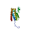
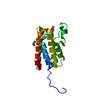
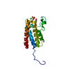
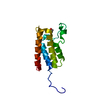
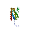
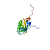
| Title | STRUCTURAL BASIS OF DIMERIZATION AND SENSORY MECHANISMS OF OXYGEN-SENSING DOMAIN OF RHIZOBIUM MELILOTI FIXL DETERMINED AT 1.4A RESOLUTION | ||||||
 Components Components | nitrogen fixation regulatory protein fixL | ||||||
 Keywords Keywords | SIGNALING PROTEIN / OXYGEN SENSOR / HISTIDINE KINASE / PAS / HIGH-RESOLUTION / TWO-COMPONENT SYSTEM | ||||||
| Function / homology |  Function and homology information Function and homology informationphosphorelay sensor kinase activity / histidine kinase / regulation of DNA-templated transcription / ATP binding / metal ion binding / plasma membrane Similarity search - Function | ||||||
| Biological species |  Sinorhizobium meliloti (bacteria) Sinorhizobium meliloti (bacteria) | ||||||
| Method |  X-RAY DIFFRACTION / SYNCHROTRON / Resolution: 1.4 Å X-RAY DIFFRACTION / SYNCHROTRON / Resolution: 1.4 Å | ||||||
 Authors Authors | Miyatake, H. / Mukai, M. / Park, S.-Y. / Adachi, S. / Tamura, K. / Nakamura, H. / Nakamura, K. / Tsuchiya, T. / Iizuka, T. / Shiro, Y. | ||||||
 Citation Citation | Journal: J.MOL.BIOL. / Year: 2000 Title: Sensory mechanism of oxygen sensor FixL from Rhizobium meliloti: crystallographic, mutagenesis and resonance Raman spectroscopic studies Authors: Miyatake, H. / Mukai, M. / Park, S.-Y. / Adachi, S. / Tamura, K. / Nakamura, H. / Nakamura, K. / Tsuchiya, T. / Iizuka, T. / Shiro, Y. | ||||||
| History |
|
- Structure visualization
| Structure viewer | Molecule: MolmilJmol/JSmol |
|---|
- Downloads & links
-Download
| PDBx/mmCIF format | 1d06.cif.gz | 41.5 KB | Display | PDBx/mmCIF format |
|---|---|---|---|---|
| PDB format | pdb1d06.ent.gz | 28 KB | Display | PDB format |
| PDBx/mmJSON format | 1d06.json.gz | Tree view | PDBx/mmJSON format | |
| Others |  Other downloads Other downloads |
-Validation report
| Arichive directory | https://data.pdbj.org/pub/pdb/validation_reports/d0/1d06ftp://data.pdbj.org/pub/pdb/validation_reports/d0/1d06 | HTTPS FTP |
|---|
-Related structure data


-Links
 PDBj
PDBj


- Assembly
| Deposited unit | 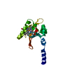
| ||||||||
|---|---|---|---|---|---|---|---|---|---|
| 1 |
| ||||||||
| 2 | 
| ||||||||
| Unit cell |
|
-Components
| #1: Protein | Mass: 14522.385 Da / Num. of mol.: 1 / Mutation: R122G, R123S, A124H, I125M, D126L, R127E Source method: isolated from a genetically manipulated source Source: (gene. exp.) Sinorhizobium meliloti (bacteria) / Plasmid: PRSET-C / Species (production host): Escherichia coli / Production host: |
|---|---|
| #2: Chemical | ChemComp-HEM /   Mass: 616.487 Da / Num. of mol.: 1 / Source method: obtained synthetically / Formula: C34H32FeN4O4 Mass: 616.487 Da / Num. of mol.: 1 / Source method: obtained synthetically / Formula: C34H32FeN4O4 |
| #3: Water | ChemComp-HOH /  Mass: 18.015 Da / Num. of mol.: 73 / Source method: isolated from a natural source / Formula: H2O Mass: 18.015 Da / Num. of mol.: 73 / Source method: isolated from a natural source / Formula: H2O |
-Experimental details
-Experiment
| Experiment | Method: X-RAY DIFFRACTION / Number of used crystals: 1 |
|---|
- Sample preparation
| Crystal | Density Matthews: 1.54 Å3/Da / Density % sol: 22 % | |||||||||||||||||||||||||
|---|---|---|---|---|---|---|---|---|---|---|---|---|---|---|---|---|---|---|---|---|---|---|---|---|---|---|
| Crystal grow | Temperature: 293 K / Method: vapor diffusion, hanging drop / pH: 4.5 Details: PEG 4000, Acetate buffer, pH 4.5, VAPOR DIFFUSION, HANGING DROP, temperature 20K | |||||||||||||||||||||||||
| Crystal grow | *PLUS Details: drop consists of equal volume of protein and reservoir solutions | |||||||||||||||||||||||||
| Components of the solutions | *PLUS
|
-Data collection
| Diffraction | Mean temperature: 100 K |
|---|---|
| Diffraction source | Source: SYNCHROTRON / Site: SPring-8  / Beamline: BL44B2 / Wavelength: 0.7 / Beamline: BL44B2 / Wavelength: 0.7 |
| Detector | Type: RIGAKU RAXIS IV / Detector: IMAGE PLATE / Date: Dec 20, 1998 / Details: MIRROR/MONOCHROMATOR |
| Radiation | Monochromator: SI(111) / Protocol: SINGLE WAVELENGTH / Monochromatic (M) / Laue (L): M / Scattering type: x-ray |
| Radiation wavelength | Wavelength: 0.7 Å / Relative weight: 1 |
| Reflection | Resolution: 1.1→100 Å / Num. all: 41962 / Num. obs: 41962 / % possible obs: 95 % / Observed criterion σ(I): 0 / Redundancy: 5.2 % / Biso Wilson estimate: 14.2 Å2 / Rmerge(I) obs: 0.087 / Net I/σ(I): 21.1 |
| Reflection shell | Resolution: 1.1→1.15 Å / Redundancy: 3 % / Rmerge(I) obs: 0.455 / Num. unique all: 890 / % possible all: 87.1 |
| Reflection | *PLUS Num. measured all: 216753 |
| Reflection shell | *PLUS % possible obs: 87.1 % |
- Processing
| Software |
| ||||||||||||||||||||||||||||||||||||
|---|---|---|---|---|---|---|---|---|---|---|---|---|---|---|---|---|---|---|---|---|---|---|---|---|---|---|---|---|---|---|---|---|---|---|---|---|---|
| Refinement | Resolution: 1.4→20 Å / Rfactor Rfree error: 0.009 / Data cutoff high absF: 10000000 / Data cutoff low absF: 0.001 / Cross valid method: THROUGHOUT / σ(F): 0 / Stereochemistry target values: Engh & Huber
| ||||||||||||||||||||||||||||||||||||
| Displacement parameters | Biso mean: 18.8 Å2 | ||||||||||||||||||||||||||||||||||||
| Refine analyze |
| ||||||||||||||||||||||||||||||||||||
| Refinement step | Cycle: LAST / Resolution: 1.4→20 Å
| ||||||||||||||||||||||||||||||||||||
| Refine LS restraints |
| ||||||||||||||||||||||||||||||||||||
| LS refinement shell | Resolution: 1.4→1.49 Å / Rfactor Rfree error: 0.026 / Total num. of bins used: 6
| ||||||||||||||||||||||||||||||||||||
| Software | *PLUS Name: X-PLOR / Version: 3.851 / Classification: refinement | ||||||||||||||||||||||||||||||||||||
| Refinement | *PLUS Rfactor Rfree: 0.278 / Rfactor Rwork: 0.221 | ||||||||||||||||||||||||||||||||||||
| Solvent computation | *PLUS | ||||||||||||||||||||||||||||||||||||
| Displacement parameters | *PLUS Biso mean: 18.46 Å2 | ||||||||||||||||||||||||||||||||||||
| Refine LS restraints | *PLUS
|
