 Movie
Movie Controller

[English] 日本語
 Yorodumi
- PDB-3lfg: Crystal structure of HPr-C-His from Thermoanaerobacter tengcongensis -
+ Open data

- Basic information
| Entry | Database: PDB / ID: 3lfg | ||||||
|---|---|---|---|---|---|---|---|
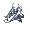
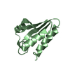
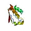
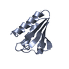
| Title | Crystal structure of HPr-C-His from Thermoanaerobacter tengcongensis | ||||||
 Components Components | Phosphotransferase system, HPr-related proteins | ||||||
 Keywords Keywords | TRANSFERASE / HPr fruB phosphotransfer PTS / Kinase / Phosphotransferase system | ||||||
| Function / homology |  Function and homology information Function and homology informationphosphoenolpyruvate-dependent sugar phosphotransferase system / cytoplasm Similarity search - Function | ||||||
| Biological species |  Thermoanaerobacter tengcongensis (bacteria) Thermoanaerobacter tengcongensis (bacteria) | ||||||
| Method |  X-RAY DIFFRACTION / SYNCHROTRON / MOLECULAR REPLACEMENT / Resolution: 1.58 Å X-RAY DIFFRACTION / SYNCHROTRON / MOLECULAR REPLACEMENT / Resolution: 1.58 Å | ||||||
 Authors Authors | Fu, T.M. / Su, X.D. | ||||||
 Citation Citation | Journal: To be Published Title: Dimerization of HPr by reversible domain swapping Authors: Fu, T.M. / Su, X.D. | ||||||
| History |
|
- Structure visualization
| Structure viewer | Molecule: MolmilJmol/JSmol |
|---|
- Downloads & links
-Download
| PDBx/mmCIF format | 3lfg.cif.gz | 115.8 KB | Display | PDBx/mmCIF format |
|---|---|---|---|---|
| PDB format | pdb3lfg.ent.gz | 91.1 KB | Display | PDB format |
| PDBx/mmJSON format | 3lfg.json.gz | Tree view | PDBx/mmJSON format | |
| Others |  Other downloads Other downloads |
-Validation report
| Arichive directory | https://data.pdbj.org/pub/pdb/validation_reports/lf/3lfgftp://data.pdbj.org/pub/pdb/validation_reports/lf/3lfg | HTTPS FTP |
|---|
-Related structure data


-Links
 PDBj
PDBj- Assembly
| Deposited unit | 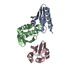
| ||||||||
|---|---|---|---|---|---|---|---|---|---|
| 1 | 
| ||||||||
| 2 | 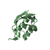
| ||||||||
| 3 | 
| ||||||||
| Unit cell |
|
-Components
| #1: Protein | Mass: 10691.167 Da / Num. of mol.: 3 Source method: isolated from a genetically manipulated source Source: (gene. exp.) Thermoanaerobacter tengcongensis (bacteria)Strain: MB4T / Gene: FruB, TTE1820 / Plasmid: pET21b / Production host: #2: Water | ChemComp-HOH / |  Mass: 18.015 Da / Num. of mol.: 275 / Source method: isolated from a natural source / Formula: H2O Mass: 18.015 Da / Num. of mol.: 275 / Source method: isolated from a natural source / Formula: H2O |
|---|
-Experimental details
-Experiment
| Experiment | Method: X-RAY DIFFRACTION / Number of used crystals: 1 |
|---|
- Sample preparation
| Crystal | Density Matthews: 2.09 Å3/Da / Density % sol: 41.01 % |
|---|---|
| Crystal grow | Temperature: 293 K / Method: vapor diffusion, sitting drop / pH: 4.6 Details: 30% PEG 4000, 0.1M NaAc pH 4.6, 0.2M ammonium acetate, VAPOR DIFFUSION, SITTING DROP, temperature 293K |
-Data collection
| Diffraction | Mean temperature: 100 K |
|---|---|
| Diffraction source | Source: SYNCHROTRON / Site: Photon Factory  / Beamline: AR-NE3A / Wavelength: 0.9798 Å / Beamline: AR-NE3A / Wavelength: 0.9798 Å |
| Detector | Type: ADSC QUANTUM 270 / Detector: CCD / Date: Jun 2, 2009 |
| Radiation | Protocol: SINGLE WAVELENGTH / Monochromatic (M) / Laue (L): M / Scattering type: x-ray |
| Radiation wavelength | Wavelength: 0.9798 Å / Relative weight: 1 |
| Reflection | Resolution: 1.58→50 Å / Num. all: 37529 / Num. obs: 37192 / % possible obs: 99.1 % / Redundancy: 7.6 % / Biso Wilson estimate: 17.36 Å2 / Rsym value: 0.116 |
| Reflection shell | Resolution: 1.58→1.61 Å / Redundancy: 4.7 % / Num. unique all: 1683 / % possible all: 91.4 |
- Processing
| Software |
| |||||||||||||||||||||||||||||||||||||||||||||||||||||||||||||||||||||||||||||||||||||||||||
|---|---|---|---|---|---|---|---|---|---|---|---|---|---|---|---|---|---|---|---|---|---|---|---|---|---|---|---|---|---|---|---|---|---|---|---|---|---|---|---|---|---|---|---|---|---|---|---|---|---|---|---|---|---|---|---|---|---|---|---|---|---|---|---|---|---|---|---|---|---|---|---|---|---|---|---|---|---|---|---|---|---|---|---|---|---|---|---|---|---|---|---|---|
| Refinement | Method to determine structure: MOLECULAR REPLACEMENT / Resolution: 1.58→28.16 Å / Occupancy max: 1 / Occupancy min: 0.3 / FOM work R set: 0.863 / SU ML: 0.23 / σ(F): 1.34 / Phase error: 19.87 / Stereochemistry target values: ML
| |||||||||||||||||||||||||||||||||||||||||||||||||||||||||||||||||||||||||||||||||||||||||||
| Solvent computation | Shrinkage radii: 0.9 Å / VDW probe radii: 1.11 Å / Solvent model: FLAT BULK SOLVENT MODEL / Bsol: 65.793 Å2 / ksol: 0.383 e/Å3 | |||||||||||||||||||||||||||||||||||||||||||||||||||||||||||||||||||||||||||||||||||||||||||
| Displacement parameters | Biso max: 62.92 Å2 / Biso mean: 19.972 Å2 / Biso min: 8.63 Å2
| |||||||||||||||||||||||||||||||||||||||||||||||||||||||||||||||||||||||||||||||||||||||||||
| Refinement step | Cycle: LAST / Resolution: 1.58→28.16 Å
| |||||||||||||||||||||||||||||||||||||||||||||||||||||||||||||||||||||||||||||||||||||||||||
| Refine LS restraints |
| |||||||||||||||||||||||||||||||||||||||||||||||||||||||||||||||||||||||||||||||||||||||||||
| LS refinement shell | Refine-ID: X-RAY DIFFRACTION / Total num. of bins used: 12
| |||||||||||||||||||||||||||||||||||||||||||||||||||||||||||||||||||||||||||||||||||||||||||
| Refinement TLS params. | Method: refined / Origin x: 11.16 Å / Origin y: -10.8294 Å / Origin z: 11.0281 Å
| |||||||||||||||||||||||||||||||||||||||||||||||||||||||||||||||||||||||||||||||||||||||||||
| Refinement TLS group |
|
