 Movie
Movie Controller


+ Open data

- Basic information
| Entry | Database: PDB / ID: 4d2k | ||||||
|---|---|---|---|---|---|---|---|
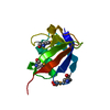
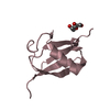
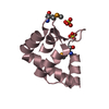
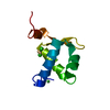
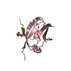
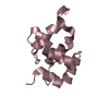
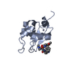
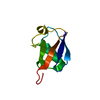
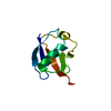
| Title | Crystal structure of DREP2 CIDE domain | ||||||
 Components Components | DREP2 | ||||||
 Keywords Keywords | APOPTOSIS / ENERGY METABOLISM / DNA FRAGMENTATION FACTOR (DFF) | ||||||
| Function / homology |  Function and homology information Function and homology informationsynaptic membrane / regulation of apoptotic process / postsynaptic membrane / apoptotic process / identical protein binding Similarity search - Function | ||||||
| Biological species |  | ||||||
| Method |  X-RAY DIFFRACTION / SYNCHROTRON / MOLECULAR REPLACEMENT / Resolution: 2.302 Å X-RAY DIFFRACTION / SYNCHROTRON / MOLECULAR REPLACEMENT / Resolution: 2.302 Å | ||||||
 Authors Authors | Jang, T.H. / Park, H.H. / Kim, Y.G. / Jeong, J.H. | ||||||
 Citation Citation | Journal: Proc. Natl. Acad. Sci. U.S.A. / Year: 2017 Title: CIDE domains form functionally important higher-order assemblies for DNA fragmentation. Authors: Choi, J.Y. / Qiao, Q. / Hong, S.H. / Kim, C.M. / Jeong, J.H. / Kim, Y.G. / Jung, Y.K. / Wu, H. / Park, H.H. | ||||||
| History |
|
- Structure visualization
| Structure viewer | Molecule: MolmilJmol/JSmol |
|---|
- Downloads & links
-Download
| PDBx/mmCIF format | 4d2k.cif.gz | 75.7 KB | Display | PDBx/mmCIF format |
|---|---|---|---|---|
| PDB format | pdb4d2k.ent.gz | 57.4 KB | Display | PDB format |
| PDBx/mmJSON format | 4d2k.json.gz | Tree view | PDBx/mmJSON format | |
| Others |  Other downloads Other downloads |
-Validation report
| Arichive directory | https://data.pdbj.org/pub/pdb/validation_reports/d2/4d2kftp://data.pdbj.org/pub/pdb/validation_reports/d2/4d2k | HTTPS FTP |
|---|
-Related structure data
| Related structure data | 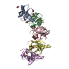 5xpcC  2eelS S: Starting model for refinement C: citing same article ( |
|---|---|
| Similar structure data |

-Links
 PDBj
PDBj
- Assembly
| Deposited unit | 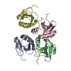
| ||||||||
|---|---|---|---|---|---|---|---|---|---|
| 1 | 
| ||||||||
| 2 | 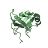
| ||||||||
| 3 | 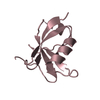
| ||||||||
| 4 | 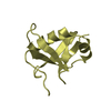
| ||||||||
| Unit cell |
|
-Components
| #1: Protein | Mass: 10063.541 Da / Num. of mol.: 4 / Fragment: CIDE DOMAIN, RESIDUES 1-84 Source method: isolated from a genetically manipulated source Source: (gene. exp.)  #2: Water | ChemComp-HOH / |  Mass: 18.015 Da / Num. of mol.: 37 / Source method: isolated from a natural source / Formula: H2O Mass: 18.015 Da / Num. of mol.: 37 / Source method: isolated from a natural source / Formula: H2O |
|---|
-Experimental details
-Experiment
| Experiment | Method: X-RAY DIFFRACTION / Number of used crystals: 1 |
|---|
- Sample preparation
| Crystal | Density Matthews: 3.44 Å3/Da / Density % sol: 64.3 % / Description: NONE |
|---|---|
| Crystal grow | pH: 6.2 Details: 100 MM BIS-TRIS PH6.2, 300 MM MAGNESIUM FORMATE DIHYDRATE |
-Data collection
| Diffraction | Mean temperature: 100 K |
|---|---|
| Diffraction source | Source: SYNCHROTRON / Site: PAL/PLS  / Beamline: 5C (4A) / Wavelength: 0.97951 / Beamline: 5C (4A) / Wavelength: 0.97951 |
| Detector | Type: ADSC QUANTUM 315r / Detector: CCD / Date: Jul 16, 2013 / Details: MIRRORS |
| Radiation | Monochromator: DOUBLE CRYSTAL MONOCHROMATOR / Protocol: SINGLE WAVELENGTH / Monochromatic (M) / Laue (L): M / Scattering type: x-ray |
| Radiation wavelength | Wavelength: 0.97951 Å / Relative weight: 1 |
| Reflection | Resolution: 2.3→50 Å / Num. obs: 22067 / % possible obs: 95.4 % / Observed criterion σ(I): 0 / Redundancy: 9.7 % / Biso Wilson estimate: 46 Å2 / Rmerge(I) obs: 0.01 / Net I/σ(I): 16.2 |
| Reflection shell | Resolution: 2.3→2.34 Å / Redundancy: 5.9 % / Rmerge(I) obs: 0.28 / Mean I/σ(I) obs: 5.2 / % possible all: 98.3 |
- Processing
| Software |
| |||||||||||||||||||||||||||||||||||||||||||||||||||||||||||||||||||||||||||||||||||||||||||||||||||||||||
|---|---|---|---|---|---|---|---|---|---|---|---|---|---|---|---|---|---|---|---|---|---|---|---|---|---|---|---|---|---|---|---|---|---|---|---|---|---|---|---|---|---|---|---|---|---|---|---|---|---|---|---|---|---|---|---|---|---|---|---|---|---|---|---|---|---|---|---|---|---|---|---|---|---|---|---|---|---|---|---|---|---|---|---|---|---|---|---|---|---|---|---|---|---|---|---|---|---|---|---|---|---|---|---|---|---|---|
| Refinement | Method to determine structure: MOLECULAR REPLACEMENT Starting model: PDB ENTRY 2EEL Resolution: 2.302→33.263 Å / SU ML: 0.29 / σ(F): 1.52 / Phase error: 24.71 / Stereochemistry target values: ML
| |||||||||||||||||||||||||||||||||||||||||||||||||||||||||||||||||||||||||||||||||||||||||||||||||||||||||
| Solvent computation | Shrinkage radii: 0.9 Å / VDW probe radii: 1.11 Å / Solvent model: FLAT BULK SOLVENT MODEL | |||||||||||||||||||||||||||||||||||||||||||||||||||||||||||||||||||||||||||||||||||||||||||||||||||||||||
| Refinement step | Cycle: LAST / Resolution: 2.302→33.263 Å
| |||||||||||||||||||||||||||||||||||||||||||||||||||||||||||||||||||||||||||||||||||||||||||||||||||||||||
| Refine LS restraints |
| |||||||||||||||||||||||||||||||||||||||||||||||||||||||||||||||||||||||||||||||||||||||||||||||||||||||||
| LS refinement shell |
|
